The nucleobase molecules carrying the genetic codes are the most important ingredients for life, but they are also very vulnerable. When the ultraviolet component in the sunlight irradiates these molecules, the electrons in the molecules will be excited, and the excited nucleobase molecules may result in irreversible changes or even damages to the DNA and RNA chains, leading to the “sunburn” of organisms at molecular level.
It is widely believed that there is a “sunscreen” mechanism in these nucleobase molecules which can lead to rapid decay into the ground state. The ultrafast decay mechanism for most types of nucleobases has been confirmed. However, the research team of Professor Todd Martinez at Stanford University proposed that there may be a shallow potential barrier for the excited electronic state of uracil (U) nucleobase, which hinders the decay of excited molecules.
This can be understood as a trick reserved by nature to promote biological variation and evolution.
This novel point of view has caused wide controversy and discussion. There are many different kinds of theoretical models about whether there is indeed a hindrance to the decay of excited state uracil. In this article, using ultrashort electron pulses and X-ray free electron lasers, the research led by Professor Zheng Li and Professor Haitan Xu provides a detailed theoretical analysis of an experimental scheme that incorporates multiple signals of ultrafast electron and X-ray diffraction and X-ray spectroscopy, and opens a way to resolve this interesting controversy.
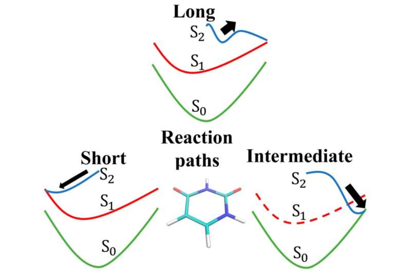
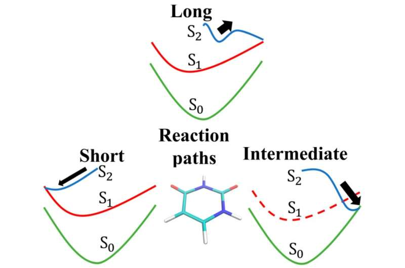
There are currently three hypotheses about the decay time scale of photoexcited uracil nucleobase. In 2007, the group of Todd Martinez proposed that the decay time of photoexcited uracil may be much longer than other nucleobases, reaching more than 1 picosecond, because the shallow potential barrier for the uracil excited state hinders the decay process.
In 2009, the research group of Zhenggang Lan from the Max Planck Institute proposed that the decay of the uracil base would not pass through the potential barrier. This theoretical model predicts short decay time of photoexcited uracil, which is about 70 femtoseconds.
In 2011, the research group of Pavel Hobza from the Institute of Organic Chemistry and Biochemistry of the Czech Academy of Sciences proposed the intermediate trajectory hypothesis, in which the uracil may have another way of structural relaxation, and the decay time through this path takes about 0.7 ps. Because the predicted potential barrier in the uracil excited state is very shallow, and due to the precision limit of quantum chemical calculations, different theoretical hypotheses give contradictory predictions of electronic decay pathways.
The authors propose an approach which can uniquely identify the electronic decay mechanism of the photoexcited uracil with ultrafast X-ray spectroscopy (XPS), ultrafast X-ray diffraction (UXD), and ultrafast electron diffraction (UED) methods. Incorporating the signatures of multiple probing methods, the authors demonstrate an approach that can identify the geometric and electronic relaxation characteristic time scales of the photoexcited uracil molecule among several candidate models.
The XPS signal provides the toolkit to map out the valence electron density variation in the chosen atomic sites of molecules. X-ray can ionize core electrons of molecules, and the shift of photoelectron energy in XPS in the molecule reflects the strength of electron screening effect of nuclear charge, which maps out the local density of valence electrons at the specific atom. Ultrafast diffraction imaging has been widely used to resolve the molecular structural dynamics.
UED is capable of characterizing the correlation between electrons and can be used to monitor the electronic population transfer dynamics. Compared to UED, UXD can resolve the transient geometric structure with higher temporal precision, which is free of pulse length limitation of UED because of space charge effect of electron bunch compression.
Combining the above signals of multiple experimental results, the characteristic time scales of geometric and electronic relaxation can be obtained, and the decay pathway of photoexcited uracil molecule can be identified.
The authors have performed molecular dynamics simulations following the long trajectory hypothesis, and calculated the ultrafast X-ray spectroscopy and coherent diffraction imaging signals. In the long trajectory hypothesis, the uracil molecule first relaxes into minimum energy geometry in the S2 state and then decays to S1 state.
The structural and electronic transition dynamics during the decay of uracil nucleobases can be reflected by XPS signal. Choosing the carbon K-edge for the X-ray probe, the variation of XPS signal in corresponding energy range is fitted, and two relaxation time scales (about 3.5 ps and 0.2 ps) are obtained.
These two characteristic time scales are related to the molecular structural evolution and electronic state transition dynamics, but the exact determination of the time scales requires combining analysis of coherent diffraction imaging, because the information of structural and electronic evolutions are usually mixed in the XPS signal.
UED is capable of characterizing the mean distance between electrons and can be used to detect the electronic population transfer dynamics. The calculated time-resolved electron diffraction signal based on molecular dynamics trajectories reflects 4.2 ps time scale of electronic state decay obtained by exponential fitting, which confirms that the 3.5 ps characteristic time scale of XPS is related to electronic transition dynamics.
The pair distribution function reflecting the average distance between atoms is obtained by Fourier transformation of UXD signal, which shows that one of the C-C bond lengths in uracil molecule is elongated in about 0.2 ps after photoexcitation followed by relaxation into minimum energy geometry in the excited state.
The time-frequency analysis of UXD signal by continuous wavelet transform reveals the frequencies of the dominant modes, and the 0.2 ps time scale of molecular structure evolution, which is consistent with the characteristic frequencies and 0.2 ps time scale of structural evolution obtained from XPS signal.
It is shown that the characteristic time scales of geometric relaxation and electronic decay of uracil in the long trajectory model can be faithfully retrieved by incorporating time-resolved XPS, UED and UXD analyses.
Incorporating the signatures of multiple probing methods, the authors demonstrate an approach to identify the decay pathway of photoexcited nucleobases among several candidate models. This study demonstrates the synergy of spectroscopic and coherent diffraction imaging with ultrafast time resolution, which can also serve as a general methodological toolkit for investigating electronic and structural dynamics in ultrafast photochemistry.
The research is published in the journal Ultrafast Science.
More information: Xiangxu Mu et al, Identification of the Decay Pathway of Photoexcited Nucleobases, Ultrafast Science (2023). DOI: 10.34133/ultrafastscience.0015
Provided by Ultrafast Science

