Tailoring fluorine-rich solid electrolyte interphase to boost long cycling stability of lithium metal batteries

Tailoring fluorine-rich solid electrolyte interphase to boost high efficiency and long cycling stability of lithium metal batter
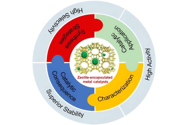
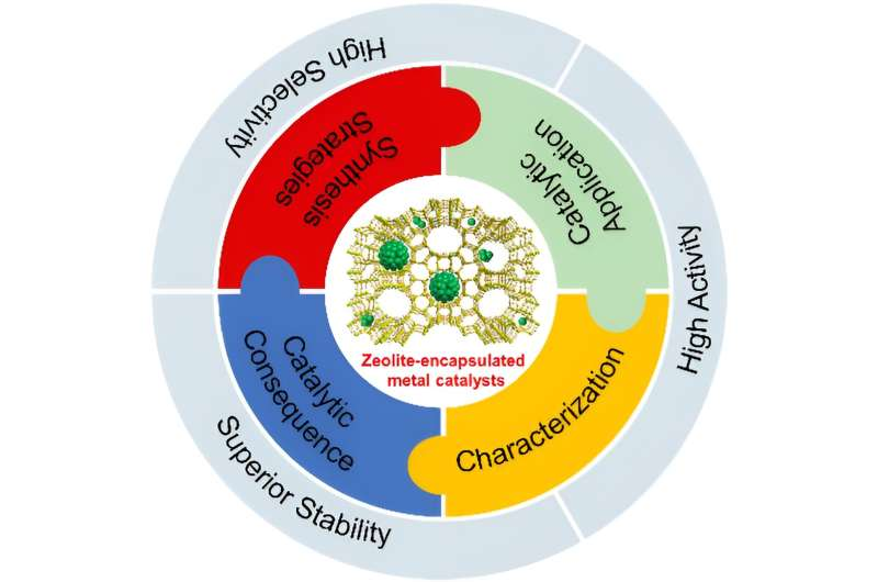
Schematics illustrating the role of DFEC in improving solid electrolyte interphase (SEI) and affection on Li deposition in ether electrolyte. Credit: Science China Press

In a study published in the journal Science China Chemistry, fluorinated cyclic carbonate (DFEC) was introduced into ether electrolyte as a SEI-forming additive. The modified electrolyte could improve the interface of Li metal anode and achieve high efficiency and long cycling stability of LMBs.

LMBs are regarded as the most promising next-generation battery system due to the high specific capacity (3860 mAh g−1) and low electrode potential (-3.04 V vs. SHE) of the Li metal anode.

However, there are many limiting factors which limit the development of LMBs—such as a side reaction between Li anode and electrolyte, Li dendrite growth and a serious volume effect of Li anode, etc.—which lead to low coulombic efficiency (CE) and poor cycle life. Stable solid electrolyte interphase (SEI) is the key to achieve high efficiency and long cycling stability of LMBs.

Adjusting SEI through electrolyte optimization is regard as a low-cost and efficient way to improve Li metal anode interface. So, it is critical to design an electrolyte formulation which can form a stable SEI, the key is the choice of solvents and film-forming additive.

Recently, Prof. Renjie Chen and Prof. Ji Qian proposed an ether-ester mixed electrolyte in which trans-difluoroethylene carbonate (DFEC) was introduced into the ether electrolyte as a film-forming additive. Firstly, ether electrolyte has good anti-reduction stability with Li metal. Secondly, due to the lower LUMO level of DFEC, it can be preferentially reduced during the initial cycle, forming LiF-rich SEI on the Li metal anode.

LiF-rich SEI can inhibit the growth of lithium dendrite, alleviate side reactions, and induce dense lithium deposition. Thanks to the above advantages, the LMBs using modified electrolyte show high efficiency and stable cycling performance. The first author of this paper is Tianyang Xue, a graduate student at Beijing Institute of Technology, and the corresponding authors are Prof. Renjie Chen, Prof. Ji Qian, and Prof. Xingming Guo.

A few implications thus emerge for designing an electrolyte to boost high efficiency and long cycling stability of LMBs. This work explores the interphase chemistry of LMBs, and provides important insights for further study on the novel electrolyte system for LMBs.

More information: Tianyang Xue et al, Tailoring fluorine-rich solid electrolyte interphase to boost high efficiency and long cycling stability of lithium metal batteries, Science China Chemistry (2023). DOI: 10.1007/s11426-022-1623-2

Provided by Science China Press 

Unraveling the role of the NiO electrocatalyst in alcohol electrooxidation reactions

Unraveling the catalytic role of NiO electrocatalyst in alcohol electrooxidation reaction
The synergy between EOM-HAT and the hydration of aldehyde results in the electrooxidation of R-CH2OH to R-COOH. The synergy between EOM-HAT and EOM-Cleavage of the C-C bond causes the electrooxidation of R-CHOH-CH2OH to R-COOH and HCOOH. Credit: Science China Press

A study led by Dr. Wei Chen, Prof. Yuqin Zou, and Prof. Shuangyin Wang (State Key Laboratory of Chemo/Bio-Sensing and Chemometrics, College of Chemistry and Chemical Engineering, Advanced Catalytic Engineering Research Center of the Ministry of Education, Hunan University) unravels the reaction mechanism of the primary alcohol/vicinal diol electrooxidation reaction on NiO, especially for the synergy between the electrochemical and non-electrochemical steps.

The alcohol electrooxidation on NiO is an indirect electrooxidation reaction with the electrophilic oxygen species as the redox mediator. Therefore, the electrochemical step is the electrochemical generation of electrophilic oxygen species. The research team found two kinds of NiO electrocatalyst function mechanisms, i.e., the electrophilic oxygen-mediated mechanism involving hydrogen atom transfer (EOM-HAT) and the electrophilic oxygen-mediated mechanism involving C-C bond cleavage (EOM-Cleavage of C-C bond).

The synergy between EOM-HAT and the hydration of aldehyde results in the electrooxidation of primary alcohol (R-CH2OH) to carboxylic acid (R-COOH) on NiO. On the other hand, the synergy between EOM-HAT and EOM-Cleavage of the C-C bond causes the electrooxidation of vicinal diol (R-CHOH-CH2OH) to R-COOH and formic acid (HCOOH) on NiO.

This study, published in the journal National Science Review, highlights the synergy between the electrochemical and non-electrochemical steps in the alcohol electrooxidation reaction. It establishes a unified reaction mechanism of alcohol electrooxidation reaction based on nickel-based electrocatalysts.

More information: Wei Chen et al, Unraveling the electrophilic oxygen-mediated mechanism for alcohol electrooxidation on NiO, National Science Review (2023). DOI: 10.1093/nsr/nwad099

Provided by Science China Press 

Mass spectrometry uncovers actions of protein ‘glues’

Mass spectrometry uncovers actions of protein 'glues'
(A) Illustrates the limited understanding of MG1 induced complex stabilization. (B) Schematic representation of MG1 ligation. (C) Enlarged view of the 14-3-3/Pin1/MG1 interface . (D) MG1 covalently bound to Lys122 of 14-3-3 by aldimine bonding. Credit: Chemical Science (2023). DOI: 10.1039/D3SC01732J

A screening technique commonly used in drug discovery can yield important details about the actions of molecular ‘glues’ in protein interactions.

Molecular glues are emerging as powerful therapeutic tools that can stick proteins together in the body. The interactions between proteins underpin all biological cell functions, including those of disease, and so interventions that can control protein-protein interactions have significant potential for disrupting the progress of various diseases.

While in many cases, drugs are required to interrupt the processes that connect proteins together, there are also occasions when the intervention is needed to restore an interaction, or to make it function correctly.

Researchers at the University of Birmingham, together with partners at the University of Leicester and the Eindhoven University of Technology, have devised a way of using mass spectrometry to analyze candidate glues for these processes and assess their relative strengths.

Dr. Aneika Leney, of the School of Biosciences at the University of Birmingham, explained, “Often when we are designing new drugs, it is to stop harmful protein interactions in the body, such as those that lead to tumor cell growth in cancers. Sometimes, however, the disease is caused by protein interactions falling apart and in these cases finding the right glue to hold them together could be extremely beneficial.”

In a new study, published in Chemical Science, the research team focused on one particular molecular glue, called MG1. Using the mass spectrometry method, they were able to disentangle the different mechanisms through which the glue bound to the proteins and stabilized the protein interaction. The MS method also allowed the researchers to elucidate the relative time taken by the different processes involved.

Dr. Peter Cossar, from the Department of Biomedical Engineering at Eindhoven University of Technology further explained, “Understanding how molecular glues stick proteins together enables scientist to better design and build the next generation of molecular glue drugs. Mass Spectrometry provides a tool to do so, by providing high fidelity information on how these unique molecules behave in real time.”

The team expect that the research will provide a robust framework for testing a wide range of molecular glues, offering a significant advance in drug discovery understanding in this area.

More information: Carlo J. A. Verhoef et al, Tracking the mechanism of covalent molecular glue stabilization using native mass spectrometry, Chemical Science (2023). DOI: 10.1039/D3SC01732J

Journal information: Chemical Science 

Provided by University of Birmingham 

Multifunctional self-healing liquid metal hydrogel developed for human-computer interaction

Multifunctional self-healing liquid metal hydrogel developed for human-computer interaction
Schematic structure and application of the liquid metal hydrogel. Credit: Li XIaofei

Recently, researchers from the Hefei Institutes of Physical Science (HFIPS) of the Chinese Academy of Sciences, led by Prof. Tian Xingyou and Prof. Zhang Xian, along with associate Prof. Yang Yanyu from the College of Materials Science and Engineering at Zhengzhou University, used gallium indium alloy (EGaIn) to initiate the polymerization and serve as flexible fillers to construct liquid metal/polyvinyl alcohol(PVA)/P(AAm-co-SMA) double network hydrogel.

“The resulting material was super-stretchable and self-healing,” said Li Xiaofei, first author of the paper, “it will promote the research and practical application of hydrogels and liquid metal in intelligent devices and military fields.”

The study was published in Materials Horizons.

Most conductive hydrogels suffer from subpar mechanical qualities and lack desirable self-recovery and self-healing abilities, severely limiting hydrogels’ potential uses. Liquid metals like gallium indium alloy (EGaIn) can toughen polymers by conforming to their changing shapes. Also, gallium (Ga) in EGaIn can initiate vinyl monomer’s free radical polymerization.

In this research, the team built a liquid metal/PVA/P(AAm-co-SMA) double network hydrogel (LM hydrogel) with EGaIn serving as both the polymerization initiator and the flexible fillers.

The PVA network used PVA microcrystals and coordination interaction of Ga3+ and PVA as cross-links, while the P(AAm-co-SMA) network used hydrophobic association and the EGaIn microspheres. The LM hydrogel was endowed with excellent super-stretchability (2000%), toughness (3.00 MJ/m3), notch resistance, and self-healing property (> 99% at 25 °C after 24 h) due to the multiple physical cross-links and the synergistic effect of the rigid PVA microcrystal network and the ductile P(AAm-co-SMA) hydrophobic network.

“The sensors developed for it can be used in health monitoring and motion identification through human-computer interaction,” said Li Xiaofei, “thanks to the LM hydrogel’s sensitive strain sensing capability.”

As a result of EGaIn’s low infrared emissivity and remarkable photothermal, LM hydrogel shows considerable promise in infrared camouflage.

More information: Xiaofei Li et al, Self-healing liquid metal hydrogel for human–computer interaction and infrared camouflage, Materials Horizons (2023). DOI: 10.1039/D3MH00341H

Journal information: Materials Horizons 

Provided by Chinese Academy of Sciences 

Stronger tape engineered through the ancient Japanese art of cutting paper, kirigami

by Virginia Tech

Stronger tape engineered through the art of cutting
Associate Professor Michael Bartlett pulls enhanced tape developed in his lab at Virginia Tech. Credit: Alex Parrish for Virginia Tech.

Adhesive tape fulfills many purposes, from quickly fixing household appliances to ensuring a reliable seal on a mailed package. When using tape with a strong bond, removing it may only be possible by scraping and prying at the tape’s corners, hoping desperately that surface pieces don’t tear away with the tape.

But what if you could make adhesives both strong and easily removable? This seemingly paradoxical combination of properties could dramatically change applications in robotic grasping, wearables for health monitoring, and manufacturing for assembly and recycling.

Developing such adhesives may not by that far off through the latest research conducted by the team of Michael Bartlett, assistant professor in the Department of Mechanical Engineering at Virginia Tech, and published in Nature Materials on June 22.

The physics of stickiness

Adhesive tapes were first developed in the 1920s to meet a need for automobile painters who wanted better options for painting two colors on car bodies. Since the first masking tape was put into use, many other variations have been created. Factories have rolled out invisible tape for wrapping presents, electrical tape for covering wires, and duct tape for more uses than it was ever intended to fill.

Associate Professor Michael Bartlett holds altered tape developed in his lab at Virginia Tech. Credit: Alex Parrish for Virginia Tech.

Normally, when tapes are peeled off, they separate in a straight line along the length of the strip until the tape is completely removed. Strong adhesives are made more difficult to peel, while reusable adhesives promote the strength-limiting separation.

Bartlett’s team theorized that if the separation path were controlled, then perhaps adhesives could be made both strong and removable. They tapped into the methods of a 2,000-year-old Japanese art form to determine how to do it.

More information: Dohgyu Hwang et al, Metamaterial adhesives for programmable adhesion through reverse crack propagation, Nature Materials (2023). DOI: 10.1038/s41563-023-01577-2 , dx.doi.org/10.1038/s41563-023-01577-2

Journal information: Nature Materials 

Provided by Virginia Tech 

Researchers discover a new way to develop drugs without side effects

A new way to develop drugs without side effects
An artist’s impression of the GPCR activation from inside the cell and the resulting targeted response. Credit: Kobayashi and Kawakami et al., 2023

Have you ever wondered how drugs reach their targets and achieve their function within our bodies? If a drug molecule or a ligand is a message, an inbox is typically a receptor in the cell membrane. One such receptor involved in relaying molecular signals is a G protein-coupled receptor (GPCR). About one-third of existing drugs work by controlling the activation of this protein. Japanese researchers now reveal a new way of activating GPCR by triggering shape changes in the intracellular region of the receptor. This new process can help researchers design drugs with fewer or no side effects.

If the cell membrane is like an Oreo cookie sandwich, GPCR is like a snake with seven segments traversing in and out of the cookie sandwich surface. The extracellular loops are the inbox for messages. When a message molecule binds to the extracellular side of the receptor, it triggers a shape change activating G proteins and the ß-arrestin protein attached to the intracellular side of the receptor. Like a molecular relay, the information passes downstream and affects various bodily processes. That is how we see, smell, and taste, which are sensations of light, smell, and taste messages.

Adverse side effects ensue if drugs acting on GPCRs activate multiple signaling pathways rather than a specific target pathway. That is why drug development focuses on activating specific molecular signal pathways within cells. Activating the GPCR from inside the cell rather than outside the cell could be one way to achieve specificity. But until now, there was no evidence of direct activation of only the intracellular side of GPCRs without the initiations from the extracellular side.

A team of researchers headed by Osamu Nureki, a professor at the University of Tokyo, and his lab, discovered a new receptor activation mode of a bone metabolism-related GPCR called human parathyroid hormone type 1 receptor (PTH1R) without signal transduction from the extracellular side.

“Understanding the molecular mechanism will enable us to design optimal drugs,” says Kazuhiro Kobayashi, a doctoral student and an author of the study. Such a drug offers “a promising treatment for osteoporosis.”

Kobayashi has been conducting research on bone formation in animal models since he was an undergrad. “Treatments for osteoporosis that target PTH1R require strict dosage, have administrative restrictions, and there aren’t yet any better alternatives,” he says. That motivated their team to look for better drug design strategies targeting the parathyroid hormone receptor.

To understand function through structure, they used cryo-electron microscopy and revealed the 3D structure of the PTH1R and G protein bound to a message molecule. The team synthesized a non-peptide message molecule called PCO371 which binds to the intracellular region of the receptor and interacts directly with G protein subunits. In other words, PCO371 activates the receptor after entering the cell.

The PCO371-bound PTH1R structure can directly and stably modulate the intracellular side of PTH1R. And because PCO371 activates only G protein and not ß-arrestin it does not cause side effects. This specificity of its binding and receptor activation mode makes it a suitable candidate for potential small-molecule-based drugs for class B1 GPCRs, like PTH1R, which currently lack oral administrative drug ligands. Such drugs would have reduced adverse effects and burdens on patients as they act on specific molecular pathways.

The findings from this study will help “develop new drugs for disorders such as obesity, pain, osteoporosis, and neurological disorders.”

The study appears in the journal Nature.

More information: Kazuhiro Kobayashi et al, Class B1 GPCR activation by an intracellular agonist, Nature (2023). DOI: 10.1038/s41586-023-06169-3

Journal information: Nature 

Provided by University of Tokyo 

Unlocking early Earth chemistry: Salt-induced changes in polyester microdroplet structure

Unlocking early Earth chemistry: Salt-induced changes in polyester microdroplet structure
Credit: Tokyo Tech

Billions of years ago, Earth was an extremely hostile planet with active volcanoes, a harsh atmosphere, and no life. This prebiotic Earth, however, was filled with a wide array of abiotic organic molecules derived from its early environment, which underwent chemical reactions that eventually led to the origin of life.

A class of such abiotic molecules abundant during the prebiotic era was the ?-hydroxy acid (?HA)–monomers with structures somewhat similar to those of the ?-amino acids essential to modern life. However, their present abundance in biology is low.

Polyester microdroplets generated from dehydration and rehydration of ?HA monomers were proposed as protocell models and could have been a type of primitive compartment that interacted with and took up various primitive analytes, such as salts within primitive aqueous environments. However, salt–polyester interactions and salt-uptake within polyester microdroplets remains poorly studied due to a lack of appropriate analytical techniques.

To bridge this gap in understanding, a team of researchers led by Special Postdoctoral Researcher Chen Chen from RIKEN (formerly of Tokyo Institute of Technology) and Specially Appointed Associate Professor Tony Z. Jia from the Earth-Life Science Institute at Tokyo Institute of Technology have recently come up with a new strategy for investigating the effect of salt uptake on polyester microdroplets.

Their breakthrough, published in Small Methods, proposed a novel way of using existing spectroscopic and biophysical methods to characterize salt uptake by polyester microdroplets and understand their salt-mediated behavior.

“Primitive molecules such as ?HAs and polyesters, though not as commonly used by current living systems as amino acids, may have laid the ground for the evolution of primitive chemical systems that led to the origin of life on Earth. Examining the interaction of polyesters with different prebiotic analytes such as salts and determining whether polyester droplets can uptake salts can provide insights into the relevant functions exhibited by primitive compartments,” explains Prof. Jia.

?HAs such as ᴅʟ-3-phenyllactic acid (PA) can undergo dehydration under early Earth mimicking conditions to form gel-like polyesters; further rehydration results in assembly of membraneless microdroplets. These membraneless droplets have previously been found to segregate primitive analytes such as nucleic acids, small organic molecules, and proteins.

Studies have hypothesized that life originated and evolved in ancient aqueous environments. If polyester microdroplets existed in primitive aqueous environments, then they might have also uptaken salts, a major analyte found in primitive aqueous environments, which could have subsequently changed the microdroplets’ structure as well.

Thus, the team subjected various ?HAs, such as PA (a neutral monomer), malic acid (a monomer with an acidic side chain), and 4-amino-2-hydroxybutyric acid (a monomer with a basic side chain) to dehydration synthesis, followed by rehydration in aqueous medium to generate neutral, acidic residue-containing, and basic residue-containing polyester microdroplets.

In fact, this study was the first to show the plausibility of acidic residue-containing polyester microdroplets! They then incubated the polyester microdroplets in aqueous solutions consisting of different concentrations of different chloride salts (NaCl, KCl, MgCl2, and CaCl2) that may have been abundant in early oceans.

Post salt uptake, the polyester microdroplets were subjected to a novel analytical technique utilizing inductively coupled plasma mass spectrometry (ICP–MS) to analyze the salt cation concentration within the microdroplets. The analyses were performed in collaboration with researchers from the Pheasant Memorial Lab at the Institute of Planetary Materials at Okayama University, where the ICP–MS was located, as part of a joint use collaborative grant.

Furthermore, in collaboration with other members, each with unique specialties, the team then coupled ICP–MS with other spectroscopic and biophysical analytical methods, such as zeta potential analysis, optical density, dynamic light scattering, and micro-Raman imaging to study in detail how salt uptake affects the surface potential, droplet turbidity, size, and internal water distribution, respectively, of the microdroplets.

The results indicated that microdroplets possessed the ability to selectively partition salt cations, leading to differential coalescence of microdroplets, likely due to reduced electrostatic repulsions between the microdroplets as a result of surface charge neutralization by the uptaken salts, which preferentially localized to the droplet surface.

The present study highlights that even slight changes in salt-uptake could significantly affect protocell structure, which could potentially account for diversity in chemistries of primitive systems that emerged in different aqueous systems—ranging from freshwater to oceanic to hypersaline under-ocean brines.

“The adoption of a novel and highly sensitive strategy for analyzing salt uptake by polyester microdroplets widened the range of known primitive chemicals that could have had an effect on primitive protocell structure and function. This opens new avenues for future investigations regarding the relevance of polyester microdroplets during the origins of life both on and off Earth,” concludes Dr. Chen.

More information: Chen Chen et al, Spectroscopic and Biophysical Methods to Determine Differential Salt‐Uptake by Primitive Membraneless Polyester Microdroplets, Small Methods (2023). DOI: 10.1002/smtd.202300119

Journal information: Small Methods 

Provided by Tokyo Institute of Technology 

Liquid metal sticks to surfaces without a binding agent

Liquid metal sticks to surfaces without a binding agent
A multifunctional Origami structure built by the liquid metal-treated paper. Credit: Cell Reports Physical Science/Yuan et al.

Everyday materials such as paper and plastic could be transformed into electronic “smart devices” by using a simple new method to apply liquid metal to surfaces, according to scientists in Beijing, China. The study, published June 9 in the journal Cell Reports Physical Science, demonstrates a technique for applying a liquid metal coating to surfaces that do not easily bond with liquid metal. The approach is designed to work at a large scale and may have applications in wearable testing platforms, flexible devices, and soft robotics.

“Before, we thought that it was impossible for liquid metal to adhere to non-wetting surfaces so easily, but here it can adhere to various surfaces only by adjusting the pressure, which is very interesting,” said Bo Yuan, a scientist at Tsinghua University and the first author of the study.

Scientists seeking to combine liquid metal with traditional materials have been impeded by liquid metal’s extremely high surface tension, which prevents it from binding with most materials, including paper. To overcome this issue, previous research has mainly focused on a technique called “transfer printing,” which involves using a third material to bind the liquid metal to the surface. But this strategy comes with drawbacks—adding more materials can complicate the process and may weaken the end product’s electrical, thermal, or mechanical performance.

To explore an alternative approach that would allow them to directly print liquid metal on substrates without sacrificing the metal’s properties, Yuan and colleagues applied two different liquid metals (eGaln and BilnSn) to various silicone and silicone polymer stamps, then applied different forces as they rubbed the stamps onto paper surfaces.

“At first, it was hard to realize stable adhesion of the liquid metal coating on the substrate,” said Yuan. “However, after a lot of trial and error, we finally had the right parameters to achieve stable, repeatable adhesion.”

The researchers found that rubbing the liquid metal-covered stamp against the paper with a small amount of force enabled the metal droplets to bind effectively to the surface, while applying larger amounts of force prevented the droplets from staying in place.

Next, the team folded the metal-coated paper into a paper crane, demonstrating that the surface can still be folded as usual after the process is completed. And after doing so, the modified paper still maintains its usual properties.

While the technique appears promising, Yuan noted that the researchers are still figuring out how to guarantee that the liquid metal coating stays in place after it has been applied. For now, a packaging material can be added to the paper’s surface, but the team hopes to figure out a solution that won’t require it.

“Just like wet ink on paper can be wiped off by hand, the liquid metal coating without packaging here also can be wiped off by the object it touches as it is applied,” said Yuan. “The properties of the coating itself will not be greatly affected, but objects in contact may be soiled.”

In the future, the team also plans to build on the method so that it can be used to apply liquid metal to a greater variety of surfaces, including metal and ceramic.

“We also plan to construct smart devices using materials treated by this method,” said Yuan.

More information: Hongzhang Wang, Direct Fabrication of Liquid Metal Multifunctional Paper Based on Force responsive Adhesion, Cell Reports Physical Science (2023). DOI: 10.1016/j.xcrp.2023.101419www.cell.com/cell-reports-phys … 2666-3864(23)00193-5

Journal information: Cell Reports Physical Science 

Provided by Cell Press 

Engineers develop a soft, printable, metal-free electrode

MIT engineers develop a soft, printable, metal-free electrode
MIT engineers developed a metal-free, Jell-O-like material that is as soft and tough as biological tissue and can conduct electricity similarly to conventional metals. The new material, which is a type of high-performance conducting polymer hydrogel, may one day replace metals in the electrodes of medical devices. Credit: Felice Frankel

Do an image search for “electronic implants,” and you’ll draw up a wide assortment of devices, from traditional pacemakers and cochlear implants to more futuristic brain and retinal microchips aimed at augmenting vision, treating depression, and restoring mobility.

Some implants are hard and bulky, while others are flexible and thin. But no matter their form and function, nearly all implants incorporate electrodes—small conductive elements that attach directly to target tissues to electrically stimulate muscles and nerves.

Implantable electrodes are predominantly made from rigid metals that are electrically conductive by nature. But over time, metals can aggravate tissues, causing scarring and inflammation that in turn can degrade an implant’s performance.

Now, MIT engineers have developed a metal-free, Jell-O-like material that is as soft and tough as biological tissue and can conduct electricity similarly to conventional metals. The material can be made into a printable ink, which the researchers patterned into flexible, rubbery electrodes. The new material, which is a type of high-performance conducting polymer hydrogel, may one day replace metals as functional, gel-based electrodes, with the look and feel of biological tissue.

“This material operates the same as metal electrodes but is made from gels that are similar to our bodies, and with similar water content,” says Hyunwoo Yuk Ph.D., co-founder of SanaHeal, a medical device startup. “It’s like an artificial tissue or nerve.”

“We believe that for the first time, we have a tough, robust, Jell-O-like electrode that can potentially replace metal to stimulate nerves and interface with the heart, brain, and other organs in the body,” adds Xuanhe Zhao, professor of mechanical engineering and of civil and environmental engineering at MIT.

Zhao, Yuk, and others at MIT and elsewhere report their results in Nature Materials. The study’s co-authors include first author and former MIT postdoc Tao Zhou, who is now an assistant professor at Penn State University, and colleagues at Jiangxi Science and Technology Normal University and Shanghai Jiao Tong University.

A true challenge

The vast majority of polymers are insulating by nature, meaning that electricity does not pass easily through them. But there exists a small and special class of polymers that can in fact pass electrons through their bulk. Some conductive polymers were first shown to exhibit high electrical conductivity in the 1970s—work that was later awarded a Nobel Prize in Chemistry.

Recently, researchers including those in Zhao’s lab have tried using conductive polymers to fabricate soft, metal-free electrodes for use in bioelectronic implants and other medical devices. These efforts have aimed to make soft yet tough, electrically conductive films and patches, primarily by mixing particles of conductive polymers, with hydrogel—a type of soft and spongy water-rich polymer.

Researchers hoped the combination of conductive polymer and hydrogel would yield a flexible, biocompatible, and electrically conductive gel. But the materials made to date were either too weak and brittle, or they exhibited poor electrical performance.

“In gel materials, the electrical and mechanical properties always fight each other,” Yuk says. “If you improve a gel’s electrical properties, you have to sacrifice mechanical properties, and vice versa. But in reality, we need both: A material should be conductive, and also stretchy and robust. That was the true challenge and the reason why people could not make conductive polymers into reliable devices entirely made out of gel.”

Electric spaghetti

In their new study, Yuk and his colleagues found they needed a new recipe to mix conductive polymers with hydrogels in a way that enhanced both the electrical and mechanical properties of the respective ingredients.

“People previously relied on homogenous, random mixing of the two materials,” Yuk says.

Such mixtures produced gels made of randomly dispersed polymer particles. The group realized that to preserve the electrical and mechanical strengths of the conductive polymer and the hydrogel respectively, both ingredients should be mixed in a way that they slightly repel—a state known as phase separation. In this slightly separated state, each ingredient could then link its respective polymers to form long, microscopic strands, while also mixing as a whole.

“Imagine we are making electrical and mechanical spaghetti,” Zhao offers. “The electrical spaghetti is the conductive polymer, which can now transmit electricity across the material because it is continuous. And the mechanical spaghetti is the hydrogel, which can transmit mechanical forces and be tough and stretchy because it is also continuous.”

The researchers then tweaked the recipe to cook the spaghettified gel into an ink, which they fed through a 3D printer, and printed onto films of pure hydrogel, in patterns similar to conventional metal electrodes.

“Because this gel is 3D-printable, we can customize geometries and shapes, which makes it easy to fabricate electrical interfaces for all kinds of organs,” says first-author Zhou.

The researchers then implanted the printed, Jell-O-like electrodes onto the heart, sciatic nerve, and spinal cord of rats. The team tested the electrodes’ electrical and mechanical performance in the animals for up to two months and found the devices remained stable throughout, with little inflammation or scarring to the surrounding tissues. The electrodes also were able to relay electrical pulses from the heart to an external monitor, as well as deliver small pulses to the sciatic nerve and spinal cord, which in turn stimulated motor activity in the associated muscles and limbs.

Going forward, Yuk envisions that an immediate application for the new material may be for people recovering from heart surgery. “These patients need a few weeks of electrical support to avoid heart attack as a side effect of surgery,” Yuk says. “So, doctors stitch a metallic electrode on the surface of the heart and stimulate it over weeks. We may replace those metal electrodes with our gel to minimize complications and side effects that people currently just accept.”

The team is working to extend the material’s lifetime and performance. Then, the gel could be used as a soft electrical interface between organs and longer-term implants, including pacemakers and deep-brain stimulators.

“The goal of our group is to replace glass, ceramic, and metal inside the body, with something like Jell-O so it’s more benign but better performance, and can last a long time,” Zhao says. “That’s our hope.”

More information: “3D Printable High Performance Conducting Polymer Hydrogel for All-Hydrogel Bioelectronic Interfaces”, Nature Materials (2023). DOI: 10.1038/s41563-023-01569-2

Journal information: Nature Materials 

Provided by Massachusetts Institute of Technology 

This story is republished courtesy of MIT News (web.mit.edu/newsoffice/), a popular site that covers news about MIT research, innovation and teaching.

Wireless sensor enables real-time spoilage alerts on food

Revolutionary sensor enables real-time spoilage alerts on food
Summary of the concept. Credit: Nature Food (2023). DOI: 10.1038/s43016-023-00750-9

Food waste and food-borne diseases are among the most critical problems urban populations face today. They contribute to greenhouse emissions tremendously and amplify economic and environmental costs. Since food spoilage remains the main reason for this waste, the circumstances of processing, transporting, and preserving food still need to be improved in line with current technological advancements.

Current monitoring processes are conducted in laboratories and use expensive chromatographic devices. These not only require too much time but also excessive resources and qualified personnel. So, present methods unfortunately prove to be inefficient in today’s circumstances.

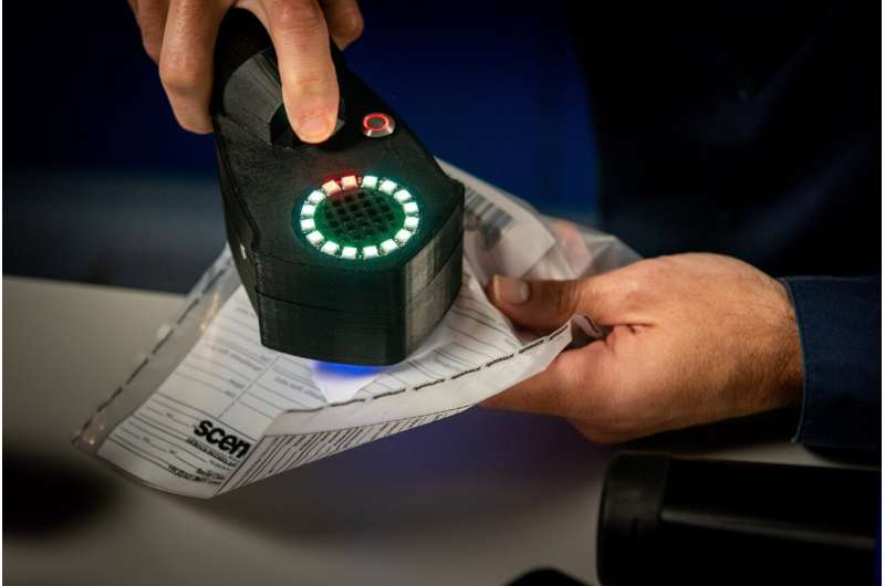
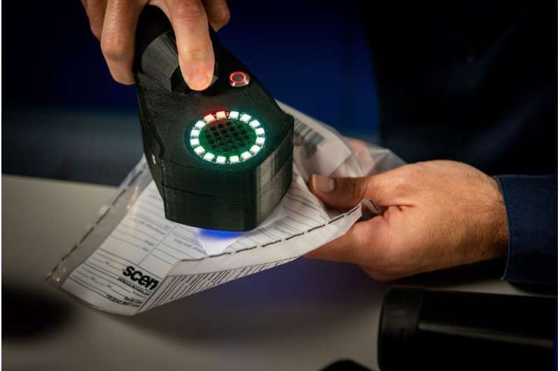
New research published in Nature Food presents a significant alternative to this process: A new user-friendly, cost-effective, and up-to-date sensor that can be applied on food directly and replace lab-monitoring. The 2 x 2 cm miniature wireless device introduced in the paper offers real-time measurement, is battery-free and smartphone-compatible. It is expected to be highly effective especially in high-protein foods such as beef, chicken, and fish.

The research was led by Dr. Emin İstif (Molecular Biology and Genetics, Kadir Has University) and Asst. Prof. Levent Beker (Mechanical Engineering, Koç University) with the contribution of Prof. İskender Yılgör and Dr. Emel Yılgör (Chemistry, Koç University), Asst. Prof. Çağdaş Dağ (Molecular Biology and Genetics, Koç University) and Asst Prof. Hatice Ceylan Koydemir (Texas A&M University).

While existing solutions focus on the change in color of food, this new device, for the first time, offers a capacitive measurement method and thus utilizes near-field communication (NFC) technology with power-free and wireless communication. The authors indicate that this eliminates major disadvantages encountered in resistive devices such as moisture sensitivity and incorrect data due to distance.

The invention will not only provide companies the opportunity of reducing costs but also help consumers tremendously. Once widely commercialized, the device will enable continuous monitoring on shelves and allow users to control freshness right before buying a product or even before consumption at home. This opportunity of on-demand spoilage analysis via mobile phones will ultimately help preventing food waste and food-borne diseases.

With its cost-effectiveness and accessibility, the authors hope to contribute to the greater struggle against global warming and greenhouse emissions more effectively and quickly. The next steps will be to focus on increasing the potential for commercialization of the product in the near future.

More information: Emin Istif et al, Miniaturized wireless sensor enables real-time monitoring of food spoilage, Nature Food (2023). DOI: 10.1038/s43016-023-00750-9

Journal information: Nature Food 

Provided by Koc University