Researchers reveal structure of unusual hormone receptor

Researchers reveal structure of unusual hormone receptor
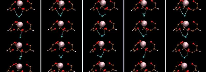
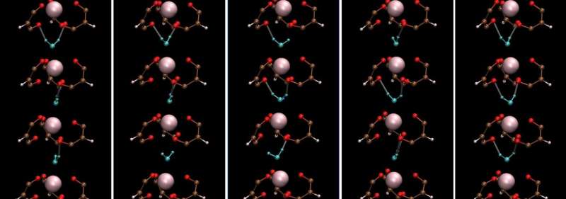
Cryo-EM formed the basis for deciphering the structure of a receptor involved in many bodily processes. Credit: Kruse lab

As the proverb goes, it takes a village to raise a child. It can also take a village to make progress in science and medicine.

In this case, labs from not one, not two, but three departments in the Blavatnik Institute at Harvard Medical School, along with a colleague in France, came together to figure out the structure of a tricky receptor involved in heart, lung, liver, and kidney function as well as pregnancy.

Untangling this structure, described April 20 in Nature Chemical Biology, provides a foundation for developing drugs that act on the receptor with the goal of treating heart disease and conditions marked by the buildup of scar tissue, or fibrosis. Those include idiopathic pulmonary fibrosis, a chronic disease of the lungs; non-alcoholic fatty liver disease; and scleroderma, which affects the skin, joints, and internal organs.

The work may also pique the interest of other scientists who study basic biology because the receptor has an unusual structure that stands out from other members of its family, the G protein-coupled receptors.

“This was a very difficult receptor to characterize,” said senior author Andrew Kruse, HMS professor of biological chemistry and molecular pharmacology. “The collaboration of multiple labs was essential, and the project highlights the value of combining experimental and computational methods.”

“You have a lot of great opportunities at HMS to work with talented scientists who use different techniques than you,” agreed Sarah Erlandson, who led the work as a graduate student in the Kruse lab. “That allowed us to form a more complete picture of the receptor and study it in more ways than we could by ourselves.”

A relaxin possibility

The receptor’s name is a mouthful: relaxin/insulin-like family peptide receptor 1, or RXFP1. It gets that name because it and its three receptor siblings bind to hormones called relaxins.

Relaxins are best known for initiating a constellation of changes in the body during pregnancy, including relaxing ligaments in the pelvis in preparation for childbirth. It also boosts sperm movement. But scientists have grown to appreciate the hormone’s many non-reproductive roles as well, including dilating blood vessels to increase blood flow, stimulating the growth of new blood vessels, breaking down collagen, and reducing inflammation and fibrosis.

Relaxins do all of this when they get released from tissues such as the heart, prostate, and placenta and bind to receptors in the membranes of cells in certain organs. The receptors then send signals that spur the cells to act.

Most relaxins stay local, but one type, relaxin-2, travels throughout the body via the blood. This is the relaxin that binds to RXFP1.

Relaxin-2’s involvement in so many bodily processes has turned scientists’ eyes toward treating diseases by mimicking higher or lower levels of the hormone. Doing so requires designing drugs that bind to RXFP1—which is hard to do without knowing its structure.

“There are no drugs available that target this receptor,” said Erlandson, who now works as a research scientist at Takeda Pharmaceuticals. “People are interested in it as an option for treatment of cardiovascular and fibrotic diseases, but when you don’t understand the detailed structure, it limits your ability to target it.”

So, the teams got to work.

It takes two to tango, four to solve a receptor structure

The Kruse lab took the first step by using cryo-electron microscopy to reveal what RXFP1 looks like at the near-atomic level when bound to relaxin-2.

But one blurry spot remained where a flexible part of the receptor kept changing position from one snapshot to the next. That elusive part was the most important one—the part that binds to relaxin.

Members of the lab of Steven Gygi, HMS professor of cell biology, tackled the problem using mass spectrometry, a different method for determining structural information that measures atomic weights. Combining the experimental cryo-EM results with the mass spectrometry data allowed Erlandson to fill in the missing structural details.

Now the researchers could see the receptor in its active, or “on,” state, bound to relaxin-2. The structure suggested that the receptor could turn itself on. If that were true, what prevented it from being on all the time, sending cell-activation signals whether relaxin was there or not?

Insight came from Debora Marks, HMS associate professor of systems biology, and Xiaojing Cong at the Institute of Functional Genomics (IGF) in France. They used computational techniques—including one called evolutionary coupling analysis, which looks at protein sequences that change together over time—to predict how different parts of the receptor might shift around between its active and inactive states.

At last, the story revealed itself.

When RXFP1 is alone, no relaxins in sight, it’s turned off. When relaxin-2 binds to it, multiple parts of the receptor change shape and communicate with one another to flip the “on” switch.

The collaboration allowed the team to answer open questions about how RXFP1’s multiple parts move around and work together to allow the receptor to do its job. The way it binds to relaxin-2 hasn’t been seen in many of its receptor relatives.

With RXFP1’s active structure in hand, researchers now have a lock to design therapeutic keys for.

“It was really exciting to get to the point in the project where we saw the structure and were making these discoveries,” said Erlandson. “There’s more work to be done, but this makes a big contribution that benefits scientists and could ultimately help patients.”

Additional authors are Shaun Rawson, James Osei-Owusu, Kelly P. Brock, Xinyue Liu, Joao A. Paulo, and Julian Mintseris, of HMS.

More information: Sarah C. Erlandson et al, The relaxin receptor RXFP1 signals through a mechanism of autoinhibition, Nature Chemical Biology (2023). DOI: 10.1038/s41589-023-01321-6

Journal information: Nature Chemical Biology 

Provided by Harvard Medical School 

Using electrified spatiotemporal heating to depolymerize plastics

Using electrified spatiotemporal heating to depolymerize plastics
The assembly of the STH system. A thinner layer of carbon felt (about 2.3 mm) is used as the top heater layer. The two ends of the top heater layer are wrapped with Cu foil electrodes for Joule heating. The top and bottom layers are placed in soft contact (without external pressure) for the STH process. A quartz tube with 10.5 mm inner diameter was used to contain the carbon bilayer structure and the reactant reservoir. The bottom image shows the heater layer exhibiting a bright orange color as we apply an electrical current through the top heater layer, demonstrating its Joule-heating capabilities. Credit: Nature (2023). DOI: 10.1038/s41586-023-05845-8

A team of engineers and materials scientists affiliated with multiple institutions in the U.S., has developed a new way to depolymerize plastics using electrified spatiotemporal heating. In their paper, published in the journal Nature, the group describes the new process and its efficiency. Nature has also published a Research Briefing in the same journal issue outlining the work done by the team.

Over the past several years, plastic pollution has become a major concern, both for the environment and for the health of plants and animals, including humans, and scientists are seeking ways to recycle it. Most of the techniques developed thus far involve using chemicals to depolymerize plastics. These efforts are still extremely inefficient, however, with yields between 10% and 25%. In this new effort, the team has found a way to use pulsed electricity to boost the yield to approximately 36%.

The approach involved designing a new kind of reactor with a porous carbon felt bilayer and a pulsed electric heater at the top. In their reactor, plastic bits are melted as they are fed in to the upper chamber and flow as a mass into a lower chamber, where the material is pushed through the felt filter. The plastic then begins to decompose as the temperature rises. As the molecules that make up the plastic become smaller, their volatility grows until they are expelled from the reactor as a gas, which allows more liquid to be drawn in. Using electricity to heat the plastic allows for oscillating the temperature, allowing simpler depolymerization reactions to take precedence over side reactions, which need additional heating to depolymerize.

In addition to improving efficiency, the new approach uses less energy because of the oscillating instead of constant heat source. The team notes the system could be made more eco-friendly by using renewable sources for the electricity. They note that their reactor does emit other materials, such as acetylene, methane and some larger molecules, along with some aromatics. They also acknowledge that more work is required to reduce the amount of carbon released during the reactions.

More information: Qi Dong et al, Depolymerization of plastics by means of electrified spatiotemporal heating, Nature (2023). DOI: 10.1038/s41586-023-05845-8

Benjamin Thompson et al, A smarter way to melt down plastics?, Nature (2023). DOI: 10.1038/d41586-023-01348-8

Journal information: Nature 

© 2023 Science X Network

Crystal phase-dependent activity of mnGaOx observed in direct syngas to light olefins

Crystal phase-dependent activity of mnGaOx observed in direct syngas to light olefins
Crystal-phase-dependent of syngas conversion activity of MnGaOx-SAPO-18. Credit: Bai Bing and Jiao Feng

Two notable metal oxide structures, spinel-type oxide and solid solution-type oxide, are widely used in Oxide-Zeolite (OXZEO) bifunctional catalysts for CO/CO2 hydrogenation reactions.

Identifying the crystallographic structure sensitivity of catalysts in chemical reactions is helpful to the rational design of catalysts. However, direct and convincing study to correlate the crystal structure of oxide to its catalytic performance has yet to be done.

Recently, a joint research team led by Prof. Bao Xinhe, Prof. Pan Xiulian, Assoc. Prof. Jiao Feng and Prof. Xiao Jianping from the Dalian Institute of Chemical Physics (DICP) of the Chinese Academy of Sciences (CAS) has observed strong crystal phase-dependent activity of MnGaOx in direct syngas conversion. This study was published in Angewandte Chemie International Edition on April 18.

“The main challenge to observing the crystal structure of oxide is the lack of a well-defined material-synthesis method to obtain metal oxides with the same element components but different crystallographic structures,” said Jiao.

In this study, the researchers found co-precipitation method and hydrothermal method could synthesize bimetallic oxides, which were composed of amorphous MnO2 and Ga2O3 with hexagonal close-packed (HCP) or Face centered-cubic (FCC) crystal phase, respectively. More interestingly, they found that the HCP oxide remained unchanged as HCP MnO-Ga2O3 solid solution oxides after reduction under H2 or CO, while the FCC solid solution oxide transformed into FCC spinel structure, where reduced Mn2+ took the A-site of AB2O4 spinel structure.

They obtained 40% CO conversion, 81% light olefins selectivity, and 0.17 g·gcat-1·h-1 space-time yield of light olefins with the combination of FCC MnGaOx-Spinel and SAPO-18. In comparison, they obtained a much inferior activity with solid solution MnGaOx with a similar chemical composition.

They further proved that the superior activity of MnGaOx-Spinel was attributed to its higher reducibility and the presence of coordinatively unsaturated Ga3+ site, which facilitated the dissociation of the C-O bond via a more efficient ketene-acetate pathway to light olefins.

“Our findings may further optimize metal oxides for OXZEO syngas conversion,” said Prof. Pan.

More information: Bing Bai et al, Tuning the Crystal Phase to Form MnGaOx‐Spinel for Highly Efficient Syngas to Light Olefins, Angewandte Chemie International Edition (2023). DOI: 10.1002/anie.202217701

Journal information: Angewandte Chemie International Edition 

Provided by Chinese Academy of Sciences 

Researchers monitor real-time single-breath exhaled hydrogen cyanide profiles

Researchers monitor real-time single-breath exhaled hydrogen cyanide profiles
Graphical abstract. Credit: Analytical Chemistry (2023). DOI: 10.1021/acs.analchem.2c05603

Hydrogen cyanide (HCN) featured with high volatility and high adsorption is a common toxic and hazardous gas. Traces of HCN are also found in human exhaled breath. Unusual high HCN concentration in the breath of cystic fibrosis (CF) patients is associated with Pseudomonas aeruginosa (PA) infection. Therefore, the development of a highly sensitive online HCN measurement in exhaled breath can enable rapid screening for PA infection in CF patients.

Recently, a research group led by Prof. Li Haiyang from the Dalian Institute of Chemical Physics (DICP) of the Chinese Academy of Sciences (CAS) has developed a flow-assisted photoionization mass spectrometry method for profiling hydrogen cyanide in exhaled breath.

The study was published in Analytical Chemistry on April 4.

“HCN is easily soluble in water and highly adsorbed on device surfaces, so sensitivity and response speed are main challenges for directly measuring HCN in exhaled breath at high humidity presents,” said Prof. Chen Ping, co-corresponding author of this study.

The researchers developed a method by utilizing a self-developed atmospheric pressure negative photoionization time-of-flight mass spectrometer instrument and improved the sensitivity and time resolution of direct HCN measurements in exhaled breath. This method enabled real-time tracking of HCN concentrations in a single exhaled breath, which could provide an effective means for early screening of CF patients with PA infection.

They proposed to use helium shield gas within the mass spectrometry ionization source. This approach reduced the effect of high humidity on ionization, improved ion transport efficiency, and thus enhanced HCN detection sensitivity.

Moreover, they improved the sampling system by shortening the sampling line and adding a gas purging process to effectively reduce HCN adsorption residue and improve time resolution. This method achieved a limit of detection of 0.3 ppbv and a resolution time of 0.5 s.

By utilizing this method, the researchers tracked the changes in the single exhaled HCN profiles of volunteers before and after gargling. The resulting profiles clearly showed an early peak and a stable end-tidal plateau, representing the concentration of the oral cavity and end-tidal gas, respectively.

“The new method demonstrated good resistance to interference and high accuracy in HCN quantification. It has potential applications in the detection of PA infection in CF patients,” said Prof. Li.

More information: Yuxuan Wen et al, Online Detection of HCN in Humid Exhaled Air by Gas Flow-Assisted Negative Photoionization Mass Spectrometry, Analytical Chemistry (2023). DOI: 10.1021/acs.analchem.2c05603

Journal information: Analytical Chemistry 

Provided by Chinese Academy of Sciences 
Researchers decipher the structure of protein methyltransferase from the mpox virus

Scientists from IOCB Prague move closer to developing a drug not only against monkeypox
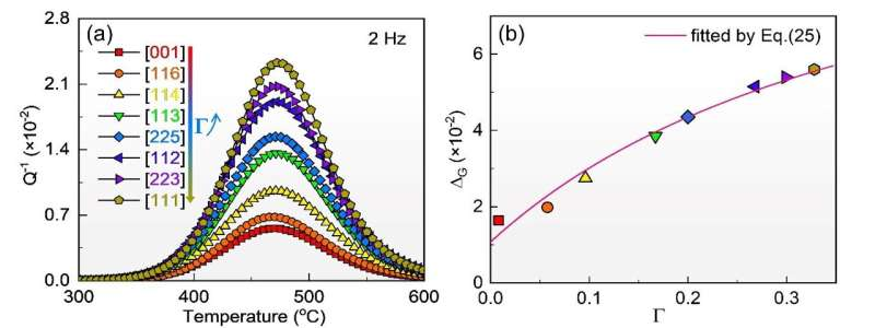
Sinefungin and SAH recognition by the monkeypox virus VP39 methyltransferase. Credit: Jan Silhan et al

Scientists from the Institute of Organic Chemistry and Biochemistry in Prague have deciphered the structure of the protein methyltransferase from the monkeypox virus. It is with the help of this protein that the virus escapes human immunity and causes the monkeypox disease. Based on this discovery, they have prepared substances that can block the function of methyltransferase.

The results of this research may constitute the first step towards creating a completely new group of antivirals. This applies not only to monkeypox, but also to diseases caused by other viruses, including COVID-19 induced by the SARS-CoV-2 coronavirus.

An article on the results of the work of the scientific groups led by Dr. Evžen Bouřa and Dr. Radim Nencka has now been published in Nature Communications. Both teams have for many years been studying viruses that cause serious diseases. In the past, they focused on the Zika virus from the flavivirus group or the SARS-CoV-2 virus from the coronavirus group.

Like other viruses, the monkeypox virus multiplies in a host cell. For it to defend itself against external attack, it needs to recognize which RNA molecules are its own and which are not. “Native RNA molecules carry a special marker called a cap for easier recognition. An unmarked molecule triggers an innate antiviral immunity response in infected cells. Therefore, viruses try to deceive the human body and, for example, the monkeypox virus confuses it by also adding a cap to its RNA,” explains Evžen Bouřa.

The symptoms of monkeypox resemble those of smallpox, a disease that has already been eradicated. Until recently, the virus causing it was found only in Central and Western Africa. Its natural reservoirs reside in rodents and primates. In humans it can cause a disease with an estimated mortality rate of 3% to 6%. While this is less than in the case of smallpox, it is much higher than, for example, with COVID-19.

Recently, the monkeypox virus has spread worldwide, so it is no wonder that not only experts, but also the general population and public authorities are nervously watching the threat of another global viral pandemic. “Our colleagues perfectly combine structural biology and cutting-edge medicinal chemistry. Thanks to that, we are closer to discovering new antivirals,” says Prof. Jan Konvalinka, the director of IOCB Prague.

More information: Jan Silhan et al, Discovery and structural characterization of monkeypox virus methyltransferase VP39 inhibitors reveal similarities to SARS-CoV-2 nsp14 methyltransferase, Nature Communications (2023). DOI: 10.1038/s41467-023-38019-1

Journal information: Nature Communications 

Provided by Institute of Organic Chemistry and Biochemistry of the CAS 

Extracting potent COVID-fighting pharmaceuticals for protective sprays

Extracting potent COVID-fighting pharmaceuticals for protective sprays
Graphical abstract. Credit: AAPS Open (2023). DOI: 10.1186/s41120-023-00072-x

David Needham, professor of mechanical engineering and materials science at Duke University, has demonstrated that a metabolic inhibiting drug called niclosamide, traditionally used to treat gut parasites, can readily be extracted and dissolved from commercial tablets in quantities sufficient to create throat and nasal sprays.

Combined with previous research that shows niclosamide might be able to prevent or inhibit the growth cycle of common respiratory viruses—including COVID-19—the results may point toward an easier pathway through the testing, regulatory approval and manufacturing processes needed to bring a potential product to market.

The research was published online April 14 in the journal American Association of Pharmaceutical Scientists Open (AAPS Open).

“These results show a potentially more rapid route to FDA approval, bringing a new commercial opportunity that could make the nasal sprays more readily available worldwide,” Needham said. “This would not just potentially be for COVID-19, but also for all respiratory viruses including Influenza and Respiratory Syncytia Virus (RSV) and to increase our preparedness for the next pandemic that seems sure to be coming down the pipe.”

Since 1958, niclosamide has been used to treat gut parasite infections in humans as well as pets and farm animals. Delivered as oral tablets, the drug kills the parasites on contact by inhibiting their crucial metabolic pathway and shutting down their energy supply.

In recent years, however, researchers have been testing niclosamide’s potential to treat a much wider range of diseases, such as many types of cancer, metabolic diseases, rheumatoid arthritis and systemic sclerosis. Recent laboratory studies in cells have also shown the drug to be a potent antiviral medication, inhibiting a virus’s ability to cause disease by targeting the energy supply of the host cell that the virus co-opts for its self-replication.

Extracting potent COVID-fighting pharmaceuticals for protective sprays
David Needham works at a lab bench to create anti-COVID throat and nasal sprays from commercial niclosamide. Credit: Duke University

Needham showed in 2022 that the drug could be dissolved into high enough concentrations to create a potential throat or nasal spray by a simple change in the solution’s pH. This was an important result, as researchers had previously believed the drug to be too insoluble to form such solutions.

Needham is now working with colleagues Zachary Kelleher in the lab of Christina Barkauskas, assistant professor of medicine in pulmonary medicine at Duke, to evaluate how niclosamide brings down the available energy in human nasal and bronchial cells. The team is also working to show that niclosamide is safe at concentrations above those where it has been found in the literature to prevent viral infection. They are now testing the effects of niclosamide in the Duke Regional Biocontainment Laboratory to see if it can prevent infection in more respiratory-relevant cells.

“Academic papers and companies actively involved in potential niclosamide products were automatically invoking much more complex formulations,” Needham said. “I showed that you could raise the solubility to what you would want for this spray-style application.”

Because the tablets are already FDA approved, the spray solution formulation seemed set for a straightforward approval and launch into a safety-efficacy clinical trial. However, the FDA viewed this throat/nasal spray as a new formulation that needed to be tested from square one, despite its concentrations being millions of times lower than the oral tablets that have been approved for more than 50 years.

By showing that enough niclosamide can be extracted from this already-approved formulation, Needham is hoping to expedite the testing and approval process.

“Having laid out the way to make the throat/nasal spray solutions by the simplest of techniques and showing that it can be readily scaled up to liters of volumes, my hope is that one or more companies will recognize not only the commercial opportunity, but also that this is the right thing to do to save lives and reduce suffering across the planet,” Needham said.

Moving forward, Needham is working to optimize these niclosamide-based solutions with an additional depot of dissolvable solid microparticles of niclosamide so that the throat and nasal tissue is continually supplied with safe concentrations of the drug. He’s also looking to engage public labs, companies, institutes and governments to make and test the formulations.

More information: David Needham, Extraction of niclosamide from commercial approved tablets into aqueous buffered solution creates potentially approvable oral and nasal sprays against COVID-19 and other respiratory infections, AAPS Open (2023). DOI: 10.1186/s41120-023-00072-x

Provided by Duke University 

Artificial photosynthesis for environmentally friendly food production

Artificial photosynthesis for environmentally friendly food production
Graphical abstract. Credit: Chem Catalysis (2023). DOI: 10.1016/j.checat.2022.100502

Ensuring the supply of food to the constantly growing world population and protecting the environment at the same time are often conflicting objectives. Now researchers at the Technical University of Munich (TUM) have successfully developed a method for the synthetic manufacture of nutritional protein using a type of artificial photosynthesis. The animal feed industry is the primary driver of high demand for large volumes of nutritional protein, which is also suitable for use in meat substitute products.

A group led by Prof. Volker Sieber at the TUM Campus Straubing for Biotechnology and Sustainability (TUMCS) has succeeded in producing the amino acid L-alanine, an essential building block in proteins, from the environmentally harmful gas CO2.

Their indirect biotechnological process involves methanol as an intermediate. Until now, protein for animal feed has been typically produced in the southern hemisphere with large-scale agricultural space requirements and negative consequences for biodiversity. The paper is published in the journal Chem Catalysis.

The CO2, which is removed from the atmosphere, is first turned into methanol using green electricity and hydrogen. The new method converts this intermediate into L-alanine in a multi-stage process using synthetic enzymes; the method is extremely effective and generates very high yields. L-alanine is one of the most important components of protein, which is essential to the nutrition of both humans and animals.

Prof. Sieber, of the TUM Professorship for Chemistry of Biogenic Resources, explains, “Compared to growing plants, this method requires far less space to create the same amount of L-alanine, when the energy used comes from solar or wind power sources. The more efficient use of space means a kind of artificial photosynthesis can be used to produce the same amount of foodstuffs on significantly fewer acres. This paves the way for a smaller ecological footprint in agriculture.”

The manufacture of L-alanine is only the first step for the scientists. “We also want to produce other amino acids from CO2 using renewable energy and to further increase efficiency in the realization process,” says co-author Vivian Willers, who developed the process as a doctoral candidate at the TUM Campus Straubing. The researchers add that the project is a good example of how bioeconomy and hydrogen economy in combination can make it possible to achieve more sustainability.

More information: Vivian Pascal Willers et al, Cell-free enzymatic L-alanine synthesis from green methanol, Chem Catalysis (2023). DOI: 10.1016/j.checat.2022.100502

Provided by Technical University Munich 

Record ammonia production achieved with inexpensive cobalt catalyst at low temperatures

Record ammonia production achieved with inexpensive cobalt catalyst at low temperatures
Credit: Tokyo Tech

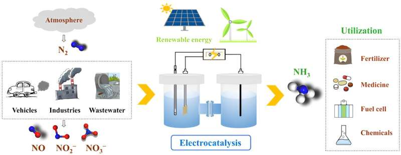
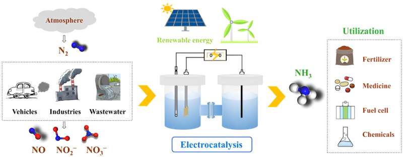
Ammonia (NH3) is one of the most widely produced chemicals in the world, with production at more than 187 million tons in 2020. About 85% of it is used to produce nitrogenous fertilizers, while the rest is used for refining petroleum, manufacturing a wide range of other chemicals, and creating synthetic fibers such as nylon. However, all this comes at a high energy cost.

Currently, most of the ammonia is produced using the conventional Haber-Bosch process, which requires combining nitrogen and hydrogen at high temperatures (400–450°C) and pressures (200 atmospheres). As a result, scientists are actively seeking catalysts that can reduce the energy requirements for ammonia production and make synthesis more sustainable.

Ruthenium (Ru), a noble metal, has been the primary candidate in this regard owing to its exceptional ability to absorb nitrogen at low temperatures. However, its high cost has prevented its widespread adoption in large-scale ammonia synthesis. While cobalt (Co) has been considered as a more cost-effective alternative, achieving the same catalytic activity as Ru at low temperatures has been difficult.

To enhance the catalytic activity of Co, a team of researchers including Professor Masaaki Kitano at Tokyo Institute of Technology (Tokyo Tech), Japan developed, in a recent study, a support material for Co nanoparticles. The material, a barium-containing oxyhydride electride called BaAl2O4-xHy, increases the catalytic activity of Co to a level comparable to that of Ru catalysts at low temperatures, and protects the H ions and electrons from the effects of air and moisture. The breakthrough was published in the Journal of the American Chemical Society.

“We attempted to develop a barium-containing oxyhydride electride, Ba2Al2O4-xHy to obtain a highly effective and chemically durable catalyst and unlock a new approach to designing novel inorganic electride materials and triggering their application in other fields,” explains Prof. Kitano.

How did the team achieve this feat? Put simply, BaAl2O4-xHy has a unique structure that promotes the dissociation of nitrogen over Co. The material exhibits a stuffed tridymite structure where AlOtetrahedra are linked to form a three-dimensional (3D) network structure, creating cage-like void spaces between the barium ions. These interstitial sites are like pockets for holding negative charges, enabling the material to donate electrons to Co and facilitate the breakdown of nitrogen molecules into nitrogen adatoms.

To improve the electron-donating ability of the material, the researchers introduced electrons to the interstitial sites by replacing the O2- lattice ions with H ions (O2- (framework)+ ½ H= H (framework) + 1/2 O2 + e (cage)). The introduction of H ions not only improved the electron-donating ability of the BaAl2Obut also facilitated the desired reduction of nitrogen to ammonia.

By promoting both the cleavage of N2 and its subsequent reduction to ammonia, the Co/Ba2Al2O4-xHcatalyst could produce more than 500 mmol of ammonia per gram of cobalt per hour, a record value for Co-based catalysts. Moreover, compared to conventional Co catalysts, which typically have activation energies for ammonia synthesis exceeding 100 kJ/mole, the proposed catalyst demonstrated an activation energy of just 48.9 kJ/mole.

Further, the stuffed tridymite structure was durable and reusable, with the AlO4-based tetrahedra framework shielding the lattice H- ions and electrons from oxidation. Finally, after exposing the Co/BaAl2O4-xHy to air, the researchers could recover up to 95% of its original activity by simply heating it in hydrogen.

With its good chemical stability, enhanced catalytic activity, and high reusability, the Co/BaAl2O4-xHy catalyst shows great promise for synthesizing ammonia at low temperatures. “This novel inorganic electride offers a new approach to developing highly effective and stable Ru-free catalysts for green ammonia synthesis,” concludes Prof. Kitano.

More information: Yihao Jiang et al, Boosted Activity of Cobalt Catalysts for Ammonia Synthesis with BaAl2O4–xHy Electrides, Journal of the American Chemical Society (2023). DOI: 10.1021/jacs.3c01074

Journal information: Journal of the American Chemical Society 

Provided by Tokyo Institute of Technology 

Birch reduction simplified to a one-minute mechanochemical process

Birch reduction simplified to a one-minute mechanochemical process
Ball milling jar and reagents used in the simplified mechanochemical Birch reduction. Credit: Koji Kubota

The traditionally cumbersome yet widely-used Birch reduction can now be carried out in a mere minute in air using an optimized mechanochemical approach.

The Birch reduction is a reaction commonly used to make medicines and bioactive compounds, but the laborious process typically requires that chemists handle liquid ammonia, use cryogenic temperatures, and carry out time-consuming steps.

Researchers at the Institute for Chemical Reaction Design and Discovery (WPI-ICReDD) in Hokkaido University have developed a simplified method for performing the Birch reduction that avoids the use of ammonia, can be done at room temperature and in ambient air, and is 20–150 times faster than conventional methods. Their findings are published in the journal Angewandte Chemie International Edition.

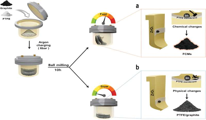
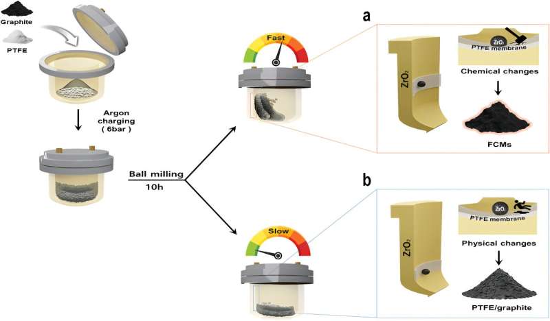
A number of lithium-based methods for performing the Birch reduction in solution have been previously developed, but since lithium reacts with both air and water, these processes still required complicated reaction setups with an inert atmosphere or dehydrated conditions. Researchers in this study saw an opportunity to avoid these issues by switching from a solution-based method to a solvent-less method using a ball mill, in which reactants are shaken rapidly in a small metal jar along with a metal ball that smashes the solid reactants together.

“In previous studies, we found that using a ball mill for reactions of metals such as magnesium and calcium with organic compounds improved the reaction rate and greatly simplified the process,” said co-author Associate Professor Koji Kubota. “Based on this, we wondered if we could develop a more straight-forward Birch reduction process by performing reactions of lithium metal with aromatic compounds in a ball mill.”

Birch reduction simplified to a one-minute mechanochemical process
Overview of the simplified protocol for the Birch reduction using a ball mill. Credit: Angewandte Chemie International Edition (2023). DOI: 10.1002/anie.202217723

The key to this strategy is that the mechanical impact from the ball breaks through the surface layer on the lithium that reacted with the air, exposing the pure lithium underneath to the other reactants and enabling the Birch reduction to proceed. This approach can be carried out in ambient air and at room temperature, making for a much easier process.

Researchers demonstrated the versatility of the process, successfully testing it with a wide variety of organic compounds, including pharmaceutical intermediates and other bioactive molecules. In most cases, the Birch reduction was completed in an astonishingly quick one minute.

The process was successfully scaled up to larger gram-scale batches, and the team believes this technique could enable the simplified synthesis of a wide variety of molecules, while also marking an important advance in mechanochemistry.

“The Birch reduction is used extensively in drug discovery and various chemical industries, and our research has made significant advancements, resulting in a much simpler and more eco-friendly Birch reduction process,” commented Professor Hajime Ito, who led the study. “We expect this breakthrough to accelerate drug discovery and various other areas of chemical research.”

More information: Yunpeng Gao et al, Mechanochemical Approach for Air‐Tolerant and Extremely Fast Lithium‐Based Birch Reductions in Minutes, Angewandte Chemie International Edition (2023). DOI: 10.1002/anie.202217723

Journal information: Angewandte Chemie International Edition 

Provided by Hokkaido University 

3D printed ferroelectric materials proven to eliminate harmful bacteria including E. coli

3D printed ferroelectric materials proven to eliminate harmful bacteria including E. coli
Schematic of the possible selective antimicrobial mechanism of the polarized 3D printed piezo-particulate PCL-BCZT composites. Credit: Advanced Materials Technologies (2023). DOI: 10.1002/admt.202202127

A new way of using 3D printing to create infection-fighting materials for use as medical implants has been revealed in a new research paper, published in Advanced Materials Technologies.

Engineers at the University of Bath, working with colleagues at the University of Ulster, have for the first time successfully created a new kind of ferroelectric composite material with antimicrobial properties using a novel multi-material 3D printing process.

They say the use of electrically responsive ferroelectric materials gives the implants the infection-fighting properties, making them ideal for biomedical applications, such as heart valves, stents and bone implants, reducing the risk of infection for patients.

Reducing risk

While commonplace, all biomedical implants pose some level of risk as materials can carry surface bio-contaminants that can lead to infection. Reducing this risk could be beneficial both to patients in the form of improved outcomes, and to healthcare providers thanks to reduced costs incurred by ongoing treatment.

The team has previously used this 3D printing technique for the fabrication of three-dimensional scaffolds for bone tissue engineering.

Dr. Hamideh Khanbareh, a lecturer in materials and structures in Bath’s Department of Mechanical Engineering, is lead author of the research. She says that the development has the scope for wide-ranging applications.

She says, “Biomedical implants that can fight infection or dangerous bacteria such as E. coli could present significant benefits to patients and to health care providers.

“Our research indicates that the ferroelectric composite materials we have created have a great potential as antimicrobial materials and surfaces. This is a potentially game-changing development that we would be keen to develop further through collaboration with medical researchers or health care providers.”

Infection-busting properties

The innovation comes thanks to ferroelectricity, a characteristic of certain polar materials that generate electrical surface charge in response to a change in mechanical energy or temperature. In ferroelectric films and implants, this electrical charge leads to the formation of free radicals known as reactive oxygen species (ROS), which selectively eradicate bacteria. This comes about through the micro-electrolysis of water molecules on a surface of polarized ferroelectric composite material.

The composite material used to harness this phenomenon is made by embedding ferroelectric barium calcium zirconate titanate (BCZT) micro-particles in polycaprolactone (PCL) a biodegradable polymer widely used in biomedical applications. The mixture of the ferroelectric particles and polymer is then fed into a 3D bioprinter to create a specific porous “scaffold” shape designed to have a high surface area to promote ROS formation.

Testing showed that even when contaminated with high concentrations of aggressive E. coli bacteria, the composite can completely eradicate the bacteria cells without external intervention, killing 70% within just 15 minutes.

More information: Zois Michail Tsikriteas et al, Additively Manufactured Ferroelectric Particulate Composites for Antimicrobial Applications, Advanced Materials Technologies (2023). DOI: 10.1002/admt.202202127

Journal information: Advanced Materials Technologies 

Provided by University of Bath