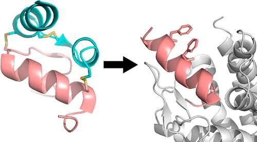
Graphical abstract. Credit: Journal of the American Chemical Society (2023). DOI: 10.1021/jacs.3c02743
In a cutting-edge discovery, published in the Journal of the American Chemical Society, Florey scientists have solved a long-standing problem: the need for an affordable, simple way to make peptide-based drugs that hold their necessary shape.
Professor Akhter Hossain, Head of the Insulin Peptides Group at The Florey, said peptides, the smaller relatives of proteins, are easy to make in a laboratory, have therapeutic potential and are considered safe. However, without the proper structure, the peptides become floppy and inactive.
“Peptides form unique structures that fit like jigsaw pieces into receptors in the brain or elsewhere in the body. They modulate a diverse range of essential bodily functions.”
Professor Hossain said our bodies naturally use complicated means of introducing structure into peptides to make them work, which is challenging to replicate in the drug development.
“Peptide stapling has been highly successful in overcoming this problem but the current methods are costly and involve complex chemistry and purification,” he said.
The paper’s lead author and Head of The Florey’s Neurotherapeutics Theme, Professor Ross Bathgate, said by simplifying peptide stapling, the team had turned a multi-step, week-long process into a shorter, single-step one.
“Our new approach is flexible, easy to implement, and will make it easier for researchers and pharmaceutical companies to develop peptide-based drugs. Now we can easily make peptides with the correct structure to bind to their target receptors. Our ultimate goal is for this technology to be used to treat a range of disorders,” Professor Bathgate said.
“In our laboratories at The Florey we’ve used this technology successfully in the earliest stage of drug discovery, and believe it will likely be applicable to a range of potential therapeutic targets for peptide-based drugs,” Professor Hossain said.
More information: Ross A. D. Bathgate et al, Noncovalent Peptide Stapling Using Alpha-Methyl-l-Phenylalanine for α-Helical Peptidomimetics, Journal of the American Chemical Society (2023). DOI: 10.1021/jacs.3c02743
Prof. Dr Ute Hellmich at the Institute for Organic Chemistry and Macromolecular Chemistry of the University Jena. Credit: Anne Günther/Uni Jena
The ordered areas of proteins are readily studied. Consequently, a great deal is known about the role of these areas in the biological function of the respective proteins. However, an international research team led by biochemist Prof. Dr. Ute Hellmich has shown that disordered areas are also pivotal.
Their comprehensive examination of the disordered area of a receptor channel protein has been published in the journal Nature Communications. The group demonstrated through eleven different methods how this area influences the function of the entire protein. Therefore, disordered protein areas should not be overlooked in research, even though they may not always be straightforward to investigate.
Investigating disorder in proteins
Proteins play a part in all processes of life. They facilitate the reading and duplication of genetic material, digest nutrients and carry out countless other essential functions. These large protein molecules can be best researched when they have a clear structure—that is, when the individual areas within the molecules are ordered.
“Classically, these proteins would be examined using X-ray crystallography or cryo-electron microscopy. But these methods are most suited for regular, or ordered, structures,” explains Hellmich. “For this reason, in some studies the disordered areas are intentionally removed to better examine the remaining molecule. But if you are specifically interested in that area, that is of course not an option.”
Hellmich and her team specifically studied a very large, disordered area of the receptor channel protein TRPV4. “So-called Transient Receptor Potential Channels, which include TRPV4, control our perception of pain and temperature and play a crucial role in the immune system and during infections,” Hellmich elucidates her research subject. “There are more than 60 known mutants of TRPV4 that cause serious illnesses. This clearly indicates how significant these proteins are,” she adds.
“In some representatives of this protein class, the disordered area comprises half of the entire molecule. This alone demonstrates that these domains cannot be overlooked,” continues the biochemist. The TRPV4 protein that her team studied has one of the largest disordered areas of this protein class in mammals, specifically containing about 130 to 150 individual amino acids. If you cut off this disordered area, TRPV4 loses its function.
A multidisciplinary perspective
In their work, the researchers used a total of eleven different biochemical and biophysical methods in various combinations—from nuclear magnetic resonance spectroscopy to mass spectrometry to molecular dynamic simulations. “At some points, we pursued a research question with different methods at the same time. We simply wanted to be quite certain that we understand the receptor correctly,” explains Hellmich. “This approach enabled us to create a molecular map of this disordered area. Thus, we discovered a network of various activity-determining elements that activate or deactivate the receptor depending on the chemical environment.
“When you consider that these disordered areas of receptor channel proteins have not been viewed in this way before, our research work certainly opens a completely new perspective on protein research and the biological function and regulation of receptors by their disordered areas,” Hellmich says. “Whether it might even be a paradigm shift, we will likely see in the coming years. I am confident that our further work within the framework of the ‘Balance of the Microverse’ excellence cluster here at the university will contribute to this,” says the scientist.
More information: Benedikt Goretzki et al, Crosstalk between regulatory elements in disordered TRPV4 N-terminus modulates lipid-dependent channel activity, Nature Communications (2023). DOI: 10.1038/s41467-023-39808-4
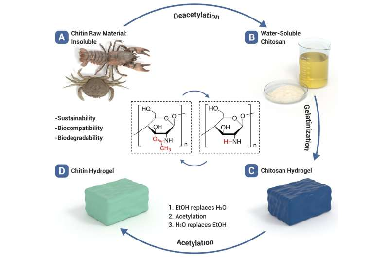
This schematic shows the preparation of chitin hydrogel via the solvent exchange-assisted acetylation of chitosan hydrogel. Credit: Nano Research, Tsinghua University Press
Chitin hydrogel is recognized as a promising material for a variety of biomedical applications. Its biocompatibility and biodegradability make it useful in tissue repair, artificial organs, and wound healing. Yet scientists continue to face challenges in fabricating chitin hydrogel. A team of researchers has developed a green, efficient and scalable preparation method for chitin hydrogels.
The team’s work provides a rational strategy to fabricate chitin hydrogels and paves the way for its practical applications as a superior biomedical material.
Their findings are published in the journal Nano Research.
Chitin, the second most abundant natural polymer, is a substance that comes from the exoskeletons of crabs, prawns, and insects. Chitin is renewable, degradable, biocompatible and low-cost. These qualities make it an excellent candidate for various biomedical uses.
“Chitin hydrogel, which shares many similarities with extracellular matrix, is an ideal material for tissue engineering and regenerative medicine. However, it is a challenge to dissolve chitin in aqueous solutions to produce hydrogel materials. Therefore, it is of great significance to develop rational fabrication strategy,” said Li-Bo Mao, a professor at the University of Science and Technology of China.
To be useful in biomedical applications, the chitin hydrogel must be biologically safe and have the appropriate mechanical strength and chemical stability. It must resist biofouling, which could lead to inflammatory response or immune rejection in the human body. For commercial use, the chitin hydrogel must also be low-cost and scalable.
The challenges in fabricating strong chitin hydrogel arise because of the insolubility of chitin in many solvents and the reduced chain length of chitin regenerated from solutions. Biopolymer hydrogels are typically prepared with a two-step process: the dissolution of the biopolymer and the subsequent gelation.
However, chitin is not soluble in water or other common solvents because of the numerous inter- and intra-molecular hydrogen bonds between the polymer chains. The team tackled this challenge by fabricating chitin hydrogel with biomimetic structure through the chemical transformation of chitosan, a water-soluble deacetylated derivative of chitin.
Chitosan is easily dissolved in water in the presence of acids. These chitosan hydrogels can be endowed with different microstructures. However, they are not mechanically or chemically stable. Attempts to improve them by using crosslinking agents have raised biosafety concerns.
The team was successful in fabricating a chemically stable and antifouling chitin hydrogel via a chemical reaction called acetylation. Through the acetylation process, the chitin hydrogel the team obtained possesses outstanding resistance to swelling, degradation, extreme temperature and pH conditions, and organic solvents.
The team also learned that by templating the chitosan precursor with ice crystals, they could produce chitin hydrogels with different biomimetic structures. These structures can be either nacre-like or wood-like depending on the freezing method used with the chitosan precursor.
The chitin hydrogel developed by the team has excellent mechanical properties while retaining a high water content. It also shows excellent antifouling performance, resisting the adhesion of proteins, bacteria, blood, and cells.
“Besides the many advantages that are characteristic to chitin, the hydrogel materials we obtained are mechanically strong and robust. In addition, the hydrogels can be feasibly processed into different shapes and structures. These ensure the practical applications of the chitin hydrogels,” said Mao.
Looking ahead, the team’s next step is to further improve the mechanical properties of chitin hydrogels and explore their biomedical applications via in vivo experiments. “We anticipate various chitin-based hydrogel materials can be fabricated through this strategy and used for different clinical applications, such as cartilage replacement, bone replacement, wound dressing and even artificial organs,” said Mao.
More information: Rui-Rui Liu et al, Biomimetic chitin hydrogel via chemical transformation, Nano Research (2023). DOI: 10.1007/s12274-023-5886-5
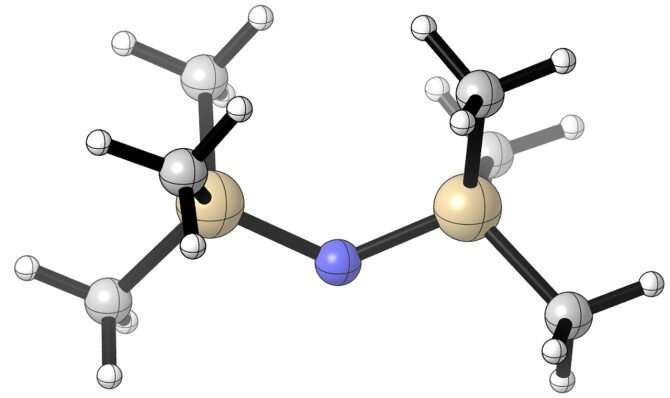
Cornell researchers bulked up highly reactive radical molecules by attaching groups of carbon and hydrogen atoms to their surface, effectively giving each molecule a set of antlers that allowed them to preserve their native reactivity while keeping their partner at a safe distance. Credit: Cornell University
How to harness the potential of highly reactive radical molecules to work in pairs and spur transformative chemistry?
Bulk them up.
That is the approach of a new Cornell-led collaboration that attached large fragments to the infamously temperamental molecules, increasing their girth to insulate them from their hyperreactive partners.
The technique could prove to be a boon for creating new and improved derivatives of pharmaceutical compounds.
The group’s paper, “Regioselective Aliphatic C–H Functionalization Using Frustrated Radical Pairs,” published July 5 in Nature. The lead author is doctoral student Zhipeng Lu.
The project, led by Song Lin, professor of chemistry and chemical biology in the College of Arts and Sciences, emerged from the Lin Group’s previous experiments with synthetic electrochemistry. In that process, electrodes pass an electrical current through a chemical reaction to activate inert molecules that will form chemical bonds that otherwise might not be achievable.
Electrochemistry also happens to be one of the most efficient ways to generate high reactive radicals from simple chemical feedstocks.
“That’s where we thought, hey, when we have these radicals, how can we control them as well? If you can harness them and use them to react with a pharmaceutical, they can do really cool chemistry,” Lin said. “It’s really our interest in electrochemistry and radical chemistry that allowed us to think about these fundamental problems.”
Radicals are highly reactive, but they have the potential to bond in pairs by sharing a pair of single electrons. The challenge is getting them close enough to cooperate without annihilating each other.
The researchers’ solution was to bulk up the radicals by attaching groups of carbon and hydrogen atoms to their surface, effectively giving each molecule a set of antlers that allowed them to preserve their native reactivity while keeping their partner at a safe distance—also known as “frustrating” them.
“We use frustrated radicals to activate carbon-hydrogen bonds and convert them into other chemical bonds, which can affect the property of the original molecule,” Lin said. “This strategy can thus be used to improve efficacy of drug molecules, for example.”
Carbon-hydrogen bonds are ideal candidates for the task because they are commonly found in organic molecules—a carbon molecule, for example, can have upwards of 20 or 30 bonds. They are also quite strong, which is why they are often conscripted into service for pharmaceutical development.
At the same time, due to their strength, carbon-hydrogen bonds can be difficult to separate out. And because they are so plentiful, selectively functionalizing individual sites is not easy.
Lin’s group collaborated with researchers from the San Francisco-based company Genentech to identify substrate targets that would enable the desired chemical reactions. The team installed large functional groups, such as trimethylsilyl, on the frustrated radicals.
“You have this barrel of substituents to make it more hindered,” Lin said. “It is actually a very simple idea. How do you use size to control the reactivity and use them to do something useful?”
Once the products were made, the team teased them apart and analyzed the reactivity and selectivity through high-level nuclear magnetic resonance and gas and liquid chromatography.
The group’s technique can help medicinal chemists initiate dozens of different chemical transformations for a range of applications, from derivatizing more efficient pharmaceutical products and enhancing their biological activity to tracking how drugs degrade in the human body.
Co-authors include postdoctoral researchers Minsoo Ju and Yi Wang; recent Cornell graduate Jonathan Meinhardt; former postdoctoral researcher Jesus Martinez Alvarado; and researchers Elisia Villemure and Jack Terrett from Genentech.
More information: Zhipeng Lu et al, Regioselective aliphatic C–H functionalization using frustrated radical pairs, Nature (2023). DOI: 10.1038/s41586-023-06131-3
Indiscriminate use of packaging materials derived from petroleum has led to a huge buildup of plastic in landfills and the ocean, as these materials have low degradability and are not significantly recycled. To mitigate this problem and meet growing demand for products that are safe for human health and the environment, the food industry is investing in the development of more sustainable packaging alternatives that preserve nutritional quality as well as organoleptic traits such as color, taste, smell and texture.
An example is a film made of a compound derived from limonene, the main component of citrus fruit peel, and chitosan, a biopolymer derived from the chitin present in exoskeletons of crustaceans.
The film was developed by a research group in São Paulo state, Brazil, comprising scientists in the Department of Materials Engineering and Bioprocesses at the State University of Campinas’s School of Chemical Engineering (FEQ-UNICAMP) and the Packaging Technology Center at the Institute of Food Technology (ITAL) of the São Paulo State Department of Agriculture and Supply, also in Campinas.
The results of the research are reported in an article published in Food Packaging and Shelf Life.
“We focused on limonene because Brazil is one of the world’s largest producers of oranges [if not the largest] and São Paulo is the leading orange-producing state,” said Roniérik Pioli Vieira, last author of the article and a professor at FEQ-UNICAMP.
Limonene has been used before in film for food packaging to enhance conservation thanks to its antioxidant and anti-microbial action, but its performance is impaired by volatility and instability during the packaging manufacturing process, even on a laboratory scale.
This is one of the obstacles to the use of bioactive compounds in commercial packaging. It is often produced in processes that involve high temperatures and high shear rates due to cutting or shaping. Bioactive additives easily degrade in these processes.
“To solve this problem, we came up with the idea of using a derivative of limonene called poly(limonene), which isn’t volatile or particularly unstable,” Vieira said.
The researchers chose chitosan for the film matrix because it is a polymer of natural origin and has well-known antioxidant and anti-microbial properties. Their hypothesis was that combining the two materials would produce a film with enhanced bioactive properties.
In the laboratory, the scientists compared films with limonene and poly(limonene) in varying proportions to address the challenge of finding a way to combine them with chitosan, since theoretically they do not mix. The researchers opted for polymerization, a process in which polymers is made from smaller organic molecules.
In this case, they used a compound with polar chemical functions to start the reaction and to increase interaction between the additive and the polymer matrix. They then analyzed the resulting film to evaluate properties such as antioxidant capacity, light and water vapor protection, and resistance to high temperatures.
The results were highly satisfactory. “The films with the poly(limonene) additive outperformed those with limonene, especially in terms of antioxidant activity, which was about twice as potent,” Vieira said. The substance also performed satisfactorily as an ultraviolet radiation blocker and was found to be non-volatile, making it suitable for large-scale production of packaging, where processing conditions are more severe.
The films are not yet available for use by manufacturers, mainly because chitosan-based plastic is not yet produced on a sufficiently large scale to be competitive, but also because the poly(limonene) production process needs to be optimized to improve yield and to be tested during the manufacturing of commercial packaging.
“Our group is working on this. We’ve investigated other applications of poly(limonene) in the biomedical field, for example. We’re trying to demonstrate the multifunctionality of this additive, whose origins are renewable,” Vieira said.
More information: Sayeny de Ávila Gonçalves et al, Poly(limonene): A novel renewable oligomeric antioxidant and UV-light blocking additive for chitosan-based films, Food Packaging and Shelf Life (2023). DOI: 10.1016/j.fpsl.2023.101085
In partnership with MoDOT, University of Missouri researchers recently created a real-world test road using recycled materials along a portion of Interstate 155 in the Missouri Bootheel. Credit: Samantha Novak/University of Missouri
Millions of roads across the United States are constructed with asphalt pavement that’s deteriorating over time. Now, researchers at the University of Missouri are using recyclables, including plastic waste, as a sustainable solution to fix America’s fracturing road system.
In partnership with the Missouri Department of Transportation (MoDOT), researchers from the Mizzou Asphalt Pavement and Innovation Lab (MAPIL) recently created a real-world test road using recycled materials like scrap tires and plastic waste along a portion of Interstate 155 in the Missouri Bootheel.
By increasing the sustainability of asphalt mixes, this innovative method can help reduce the number of items going into landfills or leaking into the environment, said Bill Buttlar, director of MAPIL.
“Missouri is the Show-Me State, so we take a very pragmatic view,” Buttlar said. “The science can be thorny and difficult, but we are up to the task. We’re excited that while our approach is complicated in the lab, its simple to execute in the field, so it makes it easily adaptable, scalable and cost-effective to incorporate into many types of road environments.”
The I-155 project takes the group’s previous test road, installed along a stretch of Stadium Boulevard in Columbia, Missouri, one step further. Instead of just testing four different types of recycled materials, the I-155 project will evaluate the real-world effectiveness of nine different types of recycled materials in the creation of asphalt pavement. This includes three different types of polyethylene (PE)—a material commonly found in plastic grocery bags—and ground tire rubber, which is a newer way of disposing scrap tires.
University of Missouri researchers are on the leading-edge of developing sustainable plastic waste road pavement mixtures. Credit: University of Missouri
“These projects afford us an opportunity to intentionally build the next generation of roads with these materials not as a type of linear landfill, but to also help the environment while making the value of dollars spent on transportation infrastructure like this stretch farther into the future,” said Buttlar, who is also the Glen Barton Chair in Flexible Pavements.
MU is on the leading-edge of this type of work in the U.S. because its team has addressed most of the translational research questions like durability and safety that could prevent a general contractor or department of transportation from adopting this ground-breaking strategy.
“We don’t just live in the laboratory,” Buttlar said. “In the field of transportation material research, we need to see how all the various materials used to construct a road—the rock, the asphalt and the recycled materials—behave in the real world and gel together to build a road.”
“Asphalt is liquefied with heat, and when you put an additive in like a plastic or rubber material, you must get everything to bond together with good adhesion. But we’re only going to know if that happens successfully when we produce it on a full-scale level and then expose it to elements, such as different weather conditions and heavy traffic.”
An asphalt pavement test mixture sample designed by the Mizzou Asphalt Pavement and Innovation Lab is ready for further lab testing to determine its strength and durability. Credit: University of Missouri
MAPIL specializes in a dry process, which allows the researchers to easily add the recyclables directly into the mixture before it’s applied to a road surface.
“The form, shape and size of the plastics bring different challenges in how the material flows, how it behaves and how it mixes,” said Punya Rath, an assistant research professor in the Department of Civil and Environmental Engineering who works at MAPIL. “So, we did extensive small-scale testing for almost an entire year before we moved to a larger scale out in the field with contractors.”
One advantage of this process is that the researchers can test the mixtures in the field using a mobile research lab, which they developed and used for both the Stadium Boulevard and I-155 projects.
“It helps the Missouri Department of Transportation (MoDOT) immensely to have a mobile research lab on-site in the field that has the ability to rapidly test samples and provide results within 24–48 hours to better inform the process,” Rath said.
Citing environmental concerns, Buttlar said the team makes sure everything they do is within the current limits as established by the Environmental Protection Agency (EPA).
“We are designing the material to be able to hold or capture the environmental by-products at the highest percentage for the longest amount of time. It’s not going to be a 100% containment,” Buttlar said. “Everything built in a natural environment will degrade over time, so that’s why EPA has standards for everything, and we make sure we are safely within those standards.”
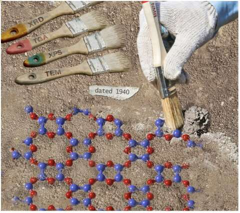
The structure of BO was a longstanding problem, dating back to 1940. Solving it required developing new NMR methods as well as applying x-ray diffraction (XRD), thermogravimetric analysis (TGA), differential scanning calorimetry (DSC), x-ray photo electron spectroscopy (XPS), and transmission electron microscopy (TEM). This research is depicted here as an archeological dig. Credit: Ames National Laboratory
In an effort to discover new 2D materials, a team of scientists from Ames National Laboratory determined the structure of boron monoxide. This compound was first discovered in the 1940s and maintained research interest throughout the years. Scientists were, however, unable to determine the structure of the material due to technological limitations of the time. Using new NMR methods and previously unavailable analytical tools, the team from Ames Lab finally solved the structure of this deceptively simple material.
“We initially weren’t really looking into studying this particular material,” said Frédéric Perras, a scientist from Ames Lab and member of the research team. “We were actually trying to make a carbon-free covalent organic framework.” A covalent organic framework is a low-density and porous material with a periodically ordered crystal structure. It is composed of organic molecules that are linked together through covalent bonds.
“However, after many synthesis trials, we could not get a highly crystalline covalent organic framework material,” said Wenyu Huang, another scientist from Ames Lab and member of the team. Perras’ and Huang’s groups are interested in these materials for alternative energy applications.
The team ended up making a boron-based material that Perras said was difficult to characterize. Through their research, they came upon literature dating back to 1940 that contained descriptions of the exact reaction the team was working on, and the synthesis of a material called boron monoxide. Unfortunately, previous scientists were unable to determine the structure of the material. This research is further discussed in the paper published in Journal of the American Chemical Society.
Luckily, technology for materials research has advanced since the 1940s. “Because of our expertise in nuclear magnetic resonance spectroscopy, and the development of new methods to which people in the 40s, 50s, and 60s didn’t have access, we thought that we might be able to lay this nearly century old mystery to rest,” said Perras.
Perras explained that boron monoxide is made using a precursor molecule that acts like building blocks. These molecules stick together through dehydration reactions. The key to understating the structure is to figure out how the blocks are physically arranged. “So we developed some NMR methods that allow us to study the orientation of these building blocks relative to each other. Basically, we found that adjacent precursor molecules were getting organized parallel to each other, which matched one of the previously proposed models,” Perras said.
“We also applied a lot of other techniques, including powder X-ray diffraction, which showed that these nanosheets organized themselves into what’s called a turbostratic arrangement,” said Perras. He explained that these stacked nanosheets are like a stack of paper thrown onto a desk. Once they land, they are not perfectly aligned, but they remain in a stack.
According to Perras, there has been a lot of recent interest in synthesizing new boron-based 2D materials. Understanding the structure of this one could lead to the synthesis of other useful boron-based 2D materials. “What really excites me is just the fact that this is an old problem. It’s such a basic material; when you write down the chemical formula, it’s BO. So, it’s interesting from that point of view that we finally solved its structure,” said Perras
More information: Frédéric A. Perras et al, The Structure of Boron Monoxide, Journal of the American Chemical Society (2023). DOI: 10.1021/jacs.3c02070
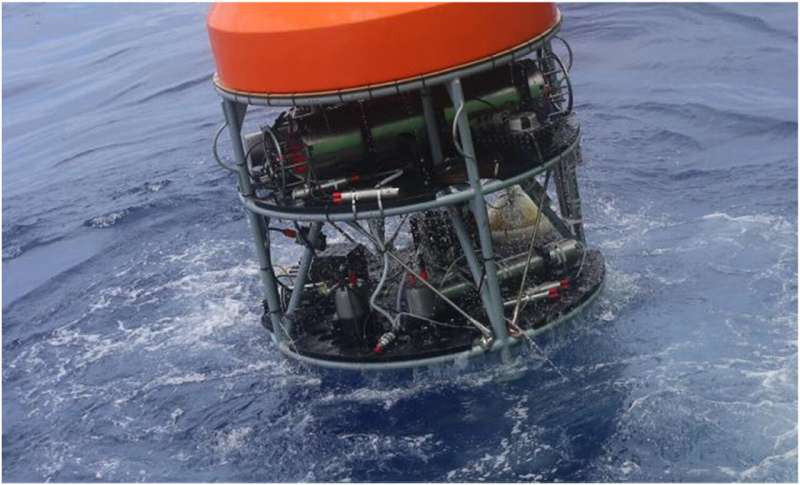
The deep-sea mass spectrometer in experiment. Credit: Wang Han
A research team led by Prof. Chen Chilai from the Hefei Institutes of Physical Science of the Chinese Academy of Sciences has developed a deep-sea mass spectrometer. It has completed several sea trials in a specific area of the deep sea.
The extreme environment of the deep sea has shaped unique biological processes and harbors significant mineral resources, the detection of which is a frontier issue in international Earth science research. Deep sea in-situ detection technology allows continuous acquisition of information on the components, concentrations and variations of deep-sea samples in both temporal and spatial dimensions. Therefore, it is increasingly being used in extreme deep-sea environments.
In this study, the deep-sea mass spectrometer worked continuously and reliably for more than eight hours under simulated water depths of -5,800 meters. It achieved long-term (25.8 hours) in-situ detection of dissolved gases in the cold seep region of the deep sea and online detection of dissolved gases from the sea surface to the seabed (-1,388 m to 0 m).
This allowed the researchers to obtain important scientific data, such as the temporal variation curve of the concentration of small molecular dissolved gases and the vertical concentration distribution profile in the deep-sea seabed. The results were published in Chinese Journal of Analytical Chemistry.
Vertical distribution of dissolved gases in the deep sea. Credit: Wang Han
This technology is not only applicable to deep-sea exploration but also to the acquisition of dissolved gas information in inland rivers, lakes and near-shore waters. Relevant research in this area is also underway.
“We have been committed to the research of key technologies and applications of mass spectrometry,” said Prof. Chen. “The development of this deep-sea mass spectrometer paves the way for the study of the origin and early evolution of life, as well as the study of global climate change.”
More information: Han Wang et al, Development and application of an underwater mass spectrometer for in situ detection of deep-sea dissolved gases, Chinese Journal of Analytical Chemistry (2023). DOI: 10.1016/j.cjac.2023.100299
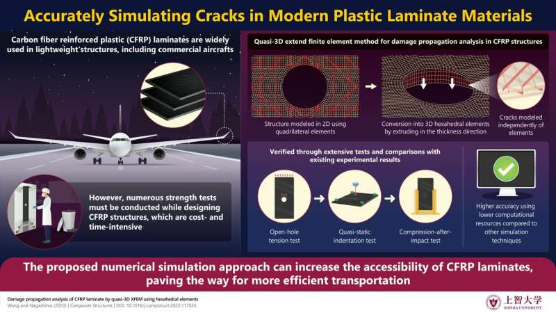
CFRPs are lightweight materials with remarkable mechanical properties. However, their design involves many time-consuming experiments, which drives up costs. To tackle this issue, researchers present a new and computationally efficient approach to simulate damage propagation in CFRP laminates. Credit: Chenyu Wang and Toshio Nagashima from Sophia University
Structural materials with useful mechanical properties have applications in a diverse range of fields. A reliable way to enhance the properties of structural materials is to make them lighter without compromising their strength.
Carbon fiber reinforced plastics (CFRPs) are perhaps the most prominent example of this approach. These plastics are made up of tiny, yet extremely strong threads of carbon atoms held together by a plastic matrix. Owing to their low weight, high durability, and exceptional mechanical performance, CFRP laminates are being incorporated into state-of-the-art aerospace applications, transportation, and construction.
However, designing CFRP laminates can be a very time-consuming endeavor. Engineers must run multiple strength tests to benchmark CFRP specimens whenever they adjust a given design. This drives up the cost of the final product and hinders the applications of CFRPs in a wider range of fields.
Against this backdrop, a duo of researchers, Dr. Chenyu Wang, a former Ph.D. student at the Graduate School of Sophia University, and Professor Toshio Nagashima from Sophia University, developed a novel method to conduct numerical simulations of damage propagation in CFRP laminates. Their findings were published online in the journal Composite Structures.
The researchers based their approach on a quasi-3D version of the extend finite element method (XFEM). In FEM, a structure or material is divided into small sub-regions known as elements, followed by the solving of physical equations for each element, to determine the overall response of the system. The “extend” version, which has been used in this study, adds functions that capture local effects around discontinuities, enabling more accurate modeling of damage propagation in form of crack growth.
Notably, since CFRP laminates are made of stacked layers of material, modeling them as flat planes (2-dimensional or 2D) would fail to capture anomalies such as delamination.
In contrast, a 3-dimensional (3D) FEM simulation would be computationally intensive and complex to set up. To overcome these issues, the researchers took a balanced approach. They first modeled the desired CFRP laminate as a 2D structure composed of quadrilateral finite elements and marked the position where cracks might occur. Then, they projected this structure in the thickness direction, while the models used to simulate delamination and matrix crack were automatically generated through their simulation system.
This strategy made the computations manageable and the modeling to simulate CFRP damage more easily and efficiently.
To verify the validity of their approach, the researchers ran simulations of three different strength and damage propagation tests on CFRP laminates and compared their results with the experimental data reported in other studies.
The first was an open-hole tension test, in which a CFRP laminate with a circular hole in the middle was pulled from one end while the other end was anchored.
The second was a quasi-static indentation (QSI) test, where a hard semi-sphere was pressed slowly and steadily against a CFRP laminate.
Lastly, the third test was the compression-after-impact test, in which the damaged specimens from the QSI test were subjected to a compressive force to assess their integrity and damage tolerance.
Overall, the results of the proposed simulation method agreed well with the experimental data, outperforming the existing quasi-3D XFEM-based techniques. Confident about the potential of the novel approach, Dr. Wang says, “The applications of composite materials such as CFRP will become more extensive and efficient if the results of this study are utilized in related fields.”
The widespread adoption of CFRPs is also likely to have important ecological implications. “In the future, if the damage of composite materials can be predicted more efficiently and accurately via numerical simulations, their cost will decrease. If these lightweight and high-strength materials are further applied in transportation, it will have a positive impact on energy savings and environmental protection,” adds Dr. Wang.
If commercial aircraft could lower their fuel consumption and time taken for design by incorporating CFRPs, the cost of flying could significantly reduce.
More information: Chenyu Wang et al, Damage propagation analysis of CFRP laminate by quasi-3D XFEM using hexahedral elements, Composite Structures (2023). DOI: 10.1016/j.compstruct.2023.117024
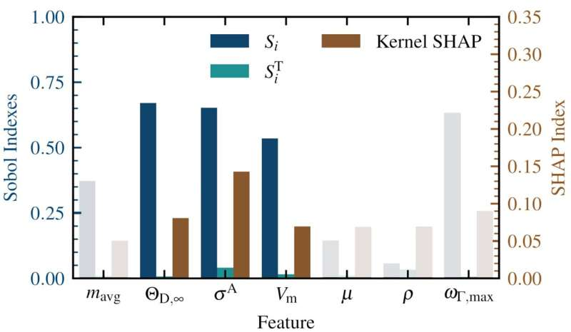
The Sobol indexes Si and SiT as well as the kernel SHAP values for each feature in the model. These values are all feature importance metrics where 0 means the feature has no effect, and a larger value means that feature is more important. Credit: Thomas Purcell
Scientists of the NOMAD Laboratory at the Fritz Haber Institute of the Max Planck Society recently proposed a workflow that can dramatically accelerate the search for novel materials with improved properties. They demonstrated the power of the approach by identifying more than 50 strongly thermally insulating materials. These can help alleviate the ongoing energy crisis, by allowing for more efficient thermoelectric elements, i.e., devices able to convert otherwise wasted heat into useful electrical voltage.
Discovering new and reliable thermoelectric materials is paramount for making use of the more than 40% of energy given off as waste heat globally and help mitigate the growing challenges of climate change. One way to increase the thermoelectric efficiency of a material is to reduce its thermal conductivity, κ, and thereby maintaining the temperature gradient needed to generate electricity.
However, the cost associated with studying these properties limited the computational and experimental investigations of κ to only a minute subset of all possible materials. A team of the NOMAD Laboratory recently made efforts to reduce these costs by creating an AI-guided workflow that hierarchically screens out materials to efficiently find new and better thermal insulators.
The work recently published in npj Computational Materials proposes a new way of using Artificial Intelligence (AI) to guide the high-throughput search for new materials. Instead of using physical/chemical intuition to screen out materials based on general, known or suspected trends, the new procedure learns the conditions that lead to the desired outcome with advanced AI methods. This work has the potential to quantify the search for new energy materials and increase the efficiency of these searches.
The first step in designing these workflows is to use advanced statistical and AI methods to approximate the target property of interest, κ in this case. To this end, the sure-independence screening and sparsifying operator (SISSO) approach is used. SISSO is a machine learning method that reveals the fundamental dependencies between different materials properties from a set of billions of possible expressions.
a) Schematic of the high-throughput workflow used to screen for new thermal insulators. b) A scatter plot showing the predicted thermal conductivity for 227 thermodynamically stable electrical insulators from both a SISSO and kernel-ridge regression (KRR) model. The color corresponds to which of the tests outlined in part a) failed. Credit: Thomas Purcell
Compared to other “black-box” AI models, this approach is similarly accurate, but additionally yields analytic relationships between different material properties. This allows us to apply modern feature importance metrics to shed light on which material properties are the most important. In the case of κ, these are the molar volume, Vm; the high-temperature limit Debye Temperature, θD,∞; and the anharmonicity metric factor, σA.
Furthermore, the described statistical analysis allows to distill out rule-of-thumbs for the individual features that enable to a priori estimate the potential of material to be a thermal insulator. Working with the three most important primary features hence allowed to create AI-guided computational workflows for discovering new thermal insulators.
These workflows use state-of-the-art electronic structure programs to calculate each of the selected features. During each step materials were screened out that are unlikely to be good insulators based on their values of Vm, θD,∞, and σA. With this, it is possible to reduce the number of calculations needed to find thermally insulating materials by over two orders of magnitude.
In this work, this is demonstrated by identifying 96 thermal insulators (κ < 10 Wm-1K-1) in an initial set of 732 materials. The reliability of this approach was further verified by calculating κ for 4 of these predictions with highest possible accuracy.
Besides facilitating the active search for new thermoelectric materials, the formalisms proposed by the NOMAD team can be also applied to solve other urgent material science problems.
More information: Thomas A. R. Purcell et al, Accelerating materials-space exploration for thermal insulators by mapping materials properties via artificial intelligence, npj Computational Materials (2023). DOI: 10.1038/s41524-023-01063-y

. DOI: 10.1021/jacs.3c02743")


, thermogravimetric analysis (TGA), differential scanning calorimetry (DSC), x-ray photo electron spectroscopy (XPS), and transmission electron microscopy (TEM). This research is depicted here as an archeological dig. Credit: Ames National Laboratory")


 Schematic of the high-throughput workflow used to screen for new thermal insulators. b) A scatter plot showing the predicted thermal conductivity for 227 thermodynamically stable electrical insulators from both a SISSO and kernel-ridge regression (KRR) model. The color corresponds to which of the tests outlined in part a) failed. Credit: Thomas Purcell")