Research and development of organic electronics such as organic solar cells and organic light-emitting diodes is rapidly advancing. The “shape” of the electron orbitals of organic molecules (molecular orbitals) is crucial to the development of organic electronics; however, methods for visualizing molecular orbitals are extremely limited.
Dynamic imaging of molecular orbitals in real space and real time has been particularly difficult yet is essential for studying structural changes and reactions of molecules.
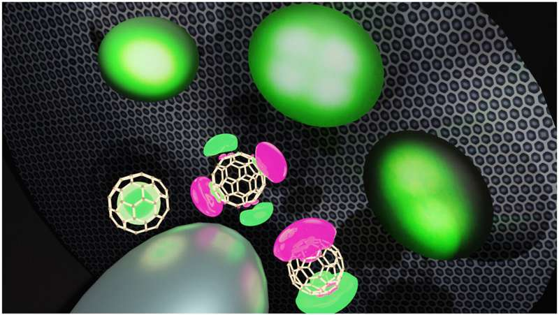
In a new study published in the journal Carbon, researchers demonstrated that the particular molecular orbitals of single molecules can be imaged by projecting the electrons emitted from organic semiconductor molecules adsorbed on a needle tip. This imaging technique is called “field emission microscopy.”
Credit: University of Tsukuba
The field emission from a molecule and its spatial distribution were analyzed in detail, revealing that the visualized orbitals might spatially extend beyond the molecule. Such orbitals, called superatom molecular orbitals (SAMOs), are suitable for electron transport in organic electronics.
These detailed measurements of SAMOs are the results of ongoing efforts by the research group. This achievement will not only facilitate future SAMO research but also promises a new dynamic method for imaging the diffusion and reactions of single molecules on surfaces.
Credit: University of Tsukuba
More information: Yoichi Yamada et al, Field emission angular distribution from single molecules, Carbon (2023). DOI: 10.1016/j.carbon.2023.118215
Credit: The Journal of Organic Chemistry (2023). DOI: 10.1021/acs.joc.3c00056
Phenanthridines are heterocyclic compounds consisting of two six-membered benzene rings fused to a six-membered ring containing nitrogen. They are found in many naturally occurring organic compounds known for their anticancer and antitumor properties. Due to their potential medicinal applications, there is a significant interest in synthesizing phenanthridine derivatives in laboratories.
A promising synthesis approach involves radical isonitrile insertion to produce imidoyl radical intermediates, which then cyclize to form phenanthridine. However, the exact mechanism of isonitrile insertion is not well understood.
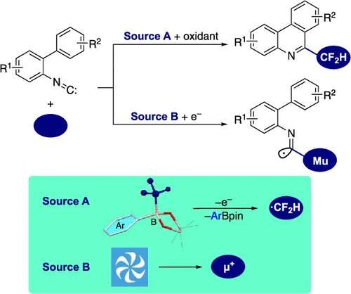
Recently, a team of researchers, led by Associate Professor Shigekazu Ito from Tokyo Institute of Technology (Tokyo Tech), has investigated the use of aryl-substituted difluoromethylborates for synthesizing difluoromethylated phenanthridines. Their study, published in The Journal of Organic Chemistry, assesses the scope of producing pharmaceutically relevant fluorinated phenanthridines from aryl-substituted difluoromethylborates and elucidates the reaction mechanism underlying isonitrile radical addition.
“Taking into account the significance of difluoromethylated phenanthridines in drug discovery, it is desirable to develop novel and complementary synthetic methods for producing 6-(difluoromethyl)phenanthridines, especially via radical isonitrile insertion,” points out Dr. Ito.
The researchers synthesized 6-(difluoromethyl)phenanthridines by first generating a highly reactive difluoromethyl radical (•CF2H) through the oxidation of aryl-substituted difluoromethylborates. This radical served as the starting point for the isonitrile insertion and cyclization processes within the isonitrile group.
After screening various oxidizing conditions, the researchers identified a combination of silver oxide (Ag2O) and potassium peroxodisulfate (K2S2O8) as ideal initiators for radical isonitrile insertion in 2-isocyano-1,1′-biphenyls. They observed that K2S2O8 oxidizes Ag2O, which, in turn, oxidizes the aryl-substituted difluoromethylborates, leading to the generation of the •CF2H radical. It attaches to the isonitrile group, producing the imidoyl radical, which then undergoes intramolecular cyclization, ultimately leading to the formation of 6-(difluoromethyl)phenanthridine.
The researchers explored various aryl groups in aryl-substituted difluoromethylborates to maximize the yield of 6-(difluoromethyl)phenanthridine. Among the tested aryl groups, p-diethylamino-phenyl-substituted borate was stable and produced the corresponding phenanthridine with a reasonable yield of 53%.
Furthermore, the researchers employed a technique called “transverse-field muon spin rotation” to confirm the reaction mechanism and the presence of the short-lived imidoyl radical. They directed a beam of positive muons (subatomic particles similar to protons but nine times lighter) towards the isonitrile group and carefully observed the changes in their spins.
Muons accompanying electrons, called muoniums, preferentially added to the carbon atom of the isonitrile unit, forming an intermediate that subsequently underwent a cyclization process. This observation provided compelling evidence for the existence of the elusive imidoyl radical.
In the future, the team hopes to explore different approaches to generate the difluoromethyl radical for facilitating the production of difluoromethylated phenanthridines. “Besides chemical oxidation, it might be possible to use photocatalytic and electrochemical methods to synthetically generate the difluoromethyl radical from difluoromethylborates,” says Dr. Ito.
In conclusion, this study presents a highly promising pathway for synthesizing 6-(difluoromethyl)phenanthridines, a breakthrough that holds tremendous potential for drug development.
More information: Kakeru Konagaya et al, Difluoromethylborates and Muonium for the Study of Isonitrile Insertion Affording Phenanthridines via Imidoyl Radicals, The Journal of Organic Chemistry (2023). DOI: 10.1021/acs.joc.3c00056
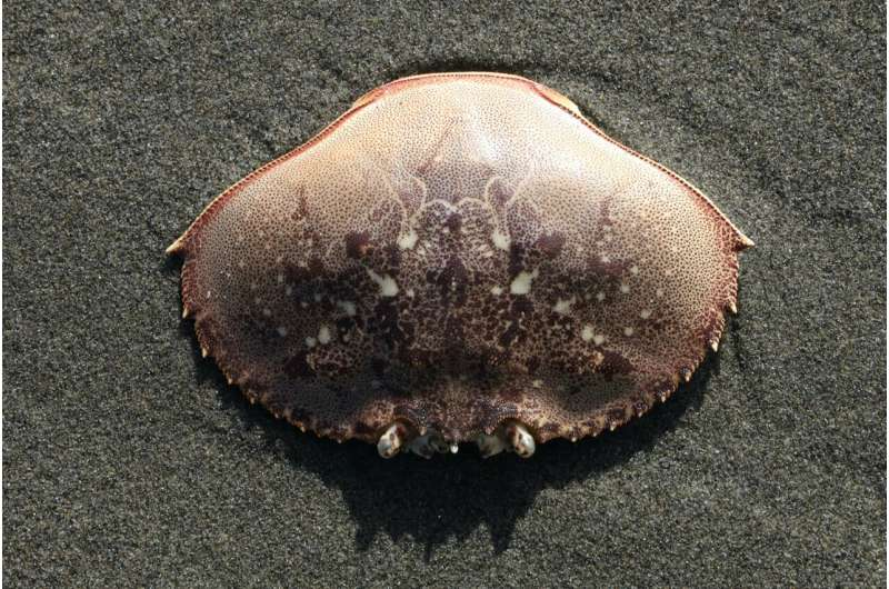
At summertime backyard feasts, crab shells are just a barrier between hunger and satisfaction. Marylanders smash the crustaceans’ protective casings with wooden mallets, pick out the tasty meat and toss the remnants aside.
But what if crab shells could have a bigger impact, playing a vital role in harnessing renewable energy and reducing planet-warming emissions?
University of Maryland researchers are changing the way people look at those thin exoskeletons—investigating the feasibility of putting them to work in an innovative battery.
“People never thought of that before,” said Lin Xu, 31, a postdoctoral researcher in the Department of Materials Science and Engineering at College Park.
Xu and a team of researchers have been exploring the use of a chemical that comes from crustacean shells in a zinc-ion battery designed to store renewable energy.
Last fall, working under the direction of Liangbing Hu, a Maryland professor who said he conceived the idea, the team published their findings on chitosan, a substance found in a variety of seafood shells, including crab and lobster.
Since appearing in a scientific journal, their work has turned heads.
“The paper has been cited already more than 20 times,” said Xu, who grew up in China and received his doctorate at Massachusetts Institute of Technology. “That’s very fast.”
He and his colleagues are attempting to solve the problem of how renewable energy—like that generated from solar or wind power—can be stored.
“It’s just like a reservoir,” Xu said of the way batteries function, essentially holding onto energy until it is needed.
At night, for example, a home’s appliances still could be powered by energy from the sun if a battery hooked up to solar panels on the roof stored energy generated during the day. On a larger scale, a battery plant placed next to a solar panel farm could stockpile energy to power a nearby city.
“We still need to find the material to store that energy, to act as a reservoir,” Xu said.
While lithium-ion batteries like those that power cellphones and electric vehicles might seem suited to the task, Xu said they are expensive, and the price tag may rise as demand grows for lithium, a finite resource.
There are also safety concerns surrounding lithium-ion batteries, which can explode and cause fires, said Xueying Zheng, a researcher who has worked alongside Xu.
“If we use a very large scale of lithium-ion batteries packed together … if one pack explodes, that will cause all of the batteries to explode,” Zheng said.
The zinc-ion battery has a different drawback: It doesn’t have a long lifespan, operating at full capacity for only a few days or a week, Xu said.
That’s where crab shells provide a solution perhaps.
With a gel membrane containing chitosan, the chemical found in seafood shells and pronounced CHI-tuh-sn, a zinc-ion battery can last a year and still function at 70% of its initial capacity. They’re also much safer, Zheng said.
The battery created and studied by UMD researchers is coin-sized, Xu said, but could be scaled up—with the goal of a more reasonable cost compared to alternatives since chitosan abounds in nature. The substance has an array of applications from biopesticides in agriculture to bandages that aid wound healing in medicine, according to Hu.
In the lab, chitosan arrives as a light yellow powder that is transformed into a translucent gel when dissolved into a solution, according to Hu, who is the director of UMD’s Center for Materials Innovation and teaches materials science and engineering.
Chitosan, a carbohydrate, “is most abundantly found in the hard outer skeletons of shellfish, including crabs, lobsters, and shrimps,” Hu wrote in an email to The Baltimore Sun. After the shells are washed and dried, they’re “pulverized into fine powders,” he explained, then treated with chemicals.
Hu’s lab has purchased chitosan from Sigma-Aldrich, a chemical and life sciences company. On its website, chitosan sells for around $300 for 250 grams, the equivalent of a little over half a pound.
A spokesperson for Merck, which owns Sigma-Aldrich, said the company could not provide details about how or where it sources chitosan since it is “proprietary information.”
“Many researchers are using our products and solutions in very interesting and unique ways,” the spokesperson told The Sun via email. “Scientific breakthroughs, both big and small, are exciting to us—especially as they positively impact life and health to create a more sustainable future.”
In Maryland, a state known for its blue crabs, some in the crab processing industry have taken notice of the potential new use for their scraps.
“I was blown away when I first saw it, thinking ‘Isn’t that crazy?'” said Jack Brooks, who read about the battery research in a seafood trade newsletter.
Brooks, 71, is president of the Chesapeake Bay Seafood Industries Association and also co-runs J.M. Clayton Co., a family-owned crab and oyster processing plant that has been operating in Cambridge since 1921.
In a single day, J.M. Clayton processes 80 to 350 bushels of crabs with each bushel containing roughly 100 crabs. The crabs are sorted and steamed before being stripped of meat in a “picking room,” Brooks explained.
From there, the discarded shells have faced different fates over the decades.
Starting in the 1920s, when J.M. Clayton operated a dehydrating plant in Cambridge, the exoskeletons were turned into a heavy powder called “crab meal,” Brooks said.
The product was used as fertilizer and chicken feed, but the equipment was “old and primitive,” he said, and his family closed the plant in the 1970s.
For about a decade after that, the shells went straight to the landfill, “which was unfortunate,” Brooks said.
Today, J.M. Clayton has a contract to provide crab shells daily—via dumpster truck—to a Dorchester County farm, where they’re used as part of a fertilizer program, he said.
“It’s a very good source of nutrients for the ground,” Brooks said.
Other area processing plants have similar arrangements, he said.
A.E. Phillips & Son, a crab processor that sells to Phillips Seafood Restaurants and other local restaurants and seafood distributors, operates a plant in Fishing Creek that has offloaded its crab shells to a farmer for use as fertilizer since 2018.
It’s the most cost-effective option for the plant, which doesn’t make any profit from the shells but likely spends less money than it would hiring a private waste removal company, said Brice Phillips, whose great-grandfather started A.E. Phillips & Son over a century ago.
“This is not just normal waste; this is waste that if you don’t get rid of it quickly, it starts to rot—and it really stinks,” said Phillips, 47, who serves as vice president of sustainability for the separate Phillips Foods.
But A.E. Phillips & Son’s processing of 60,000 pounds of crab meat per year in Maryland is dwarfed by Phillips Foods’ production in Asia. There, Phillips said, four factories in Indonesia, one in Vietnam and another in India process a combined 100,000 pounds of crab meat each week.
Phillips said he’s not sure what happens to the crab shells after they’re picked at those plants. But he suggested Asia is an ideal place for innovation.
“Whoever’s running this battery research, if they’re ever going to do anything with this, they’re basically going to be setting up a plant in Asia to get the crab shells,” Phillips said.
In Asia, each pound of crab meat comes with four pounds of “guts and shells,” he noted.
Both Brooks and Phillips said they’d be open to embracing a new use for shells.
“We’ve seen ideas come and go, but in this day and time, with all the research and technology and creative minds out there, I mean, hey, anything’s possible,” Brooks said.
Phillips views it as a potentially fruitful business venture, especially since “it seems there is no demand” for crab shells currently.
“My entrepreneurial spirit’s already just grinding the gears, trying to figure out what’s the best way to collect this stuff in mass,” he mused. “How would it be processed, where would it be processed? Where would the battery production be?”
There’s still a long way to go to make chitosan-based batteries a reality outside of the lab. A startup to commercialize the new technology is in its infancy, according to Xu.
If chitosan proves to be part of the solution—and if locally processed crab shells can be put to use—it’s likely something people in the state would get behind.
“Marylanders certainly love their crabs, and I think most people like renewable energy,” Phillips said.
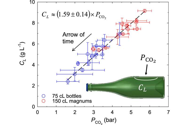
Tiny bubbles bursting in a drinker’s face and the bite of carbonation are all part of the experience when sipping champagne and sparkling wines. But how long can these drinks be stored in sealed bottles before they go flat? According to researchers reporting in ACS Omega, the answer depends on the container’s size. They estimate a 40-year shelf-life for 750-milliliter (25-ounce) bottles, and 82 and 132 years for 1.5-liter (50-ounce) and 3-liter (101-ounce) bottles, respectively.
Champagne and other sparkling wines get their bubbliness and tingly sensation from carbon dioxide, which is generated during a second round of fermentation that happens inside their bottles. Combining yeasts, sugar and wine launches the production of this gas and additional alcohol. Although the yeast die within a few months, complex aromas develop as the bottles age undisturbed for 15 months to several decades. But at the same time, the beverage is losing carbon dioxide, which is slowly escaping through the sealed metal caps or corks. So, Gérard Liger-Belair and colleagues wanted to answer the question: How does the size of the bottle influence how long you can age a champagne before it’s flat?
The researchers measured the carbon dioxide in different champagne vintages aged for multiple decades, and estimated the original amount of yeast-produced carbon dioxide. They found that the amount of gas inside the vessels, which were sealed with metal caps, decreased the longer the bottles aged. For example, the oldest vintage from 1974 lost the most carbonation, nearly 80%. Additionally, the team observed a correlation between the volume of a bottle and the carbon dioxide level, such that larger bottles retained gas substantially better than smaller ones.
In the end, the researchers developed a formula to calculate a bottle’s shelf life, or how long aged champagne would still spontaneously produce bubbles when poured in a glass. They predicted a shelf life of 40 years for standard 750-milliliter bottles, 82 years for 1.5-liter bottles and 132 years for 3-liter bottles, after which point the champagne would be flat. From their large selection of aged champagne, going back nearly 50 years, the researchers say they’ve shown how the drink’s bubbliness over time depends on the bottle’s size.
More information: Gérard Liger-Belair et al, Losses of Yeast-Fermented Carbon Dioxide during Prolonged Champagne Aging: Yes, the Bottle Size Does Matter!, ACS Omega (2023). DOI: 10.1021/acsomega.3c01812
More than 20 enzymes are involved in the production of the 21R-citrinadin A molecule. Credit: Gustavo Raskosky/Rice University
Many of the drugs we use to treat cancer and infectious disease are—or derive from— natural products, but it’s difficult to know exactly how nature assembles them.
Retracing nature’s steps, Rice University chemical engineer Xue Gao and her team mapped out the full series of enzyme-powered reactions a marine fungus uses to produce 21R-citrinadin A, a complex molecule with anticancer properties.
In the process, Gao and her collaborators identified a new enzyme, CtdY, which is the only one of its kind known to break an amide bond, according to the new study published in the Journal of the American Chemical Society.
“CtdY belongs to a large family of enzymes known as cytochrome P450s that perform a variety of different functions and are being studied for their potential use in industrial and pharmaceutical settings,” Gao said. “However, none of the P450s documented so far can break an amide bond.
“Amide bonds are found in all proteins—they’re the ones linking the amino acids together. It’s a fundamental, very stable type of bond.”
The enzyme’s ability to cleave amide bonds could make it a useful tool for creating new drugs.
“The fact that CtdY can do this is quite remarkable,” said Qiuyue Nie, a postdoctoral researcher in the Gao lab who is one of the lead authors of the study. “It holds significant promise for the pharmaceutical industry,” she said.
Qiuyue Nie (left) and Shuai Liu are lead co-authors on the study published in the Journal of the American Chemical Society. Credit: Gustavo Raskosky/Rice University
The enzyme is notable not only because it can break a highly-stable bond, but also because it does so for a very complex molecular structure.
“You want to maintain the rest of this structure and only want to break this single, hard-to-break bond ⎯ this is a very specific and difficult task,” Gao said.
Once CtdY breaks the amide bond—which has a circular 3D structure—a group of seven other enzymes intervene to complete the assembly of the 21R-citrinadin A molecule.
“Once it opens the ring, all the other enzymes are able to perform oxidation and install oxygen-hydrogen groups in a highly precise way,” Gao said. “It’s like CtdY brings the Christmas tree home, and then these other enzymes come together to decorate it.”
The Gao lab has been working for years to uncover all the steps involved in the production of the 21R-citrinadin A compound, which has been shown to be effective against leukemia in rats and human throat cancer cells, according to Shuai Liu, a Rice postdoctoral researcher who is the study’s lead co-author.
The newly identified enzyme is one of several discovered by the Gao lab that can perform singular catalytic functions such as controlling chirality and facilitating the Diels-Alder reaction.
“This really is a complete story,” Gao said. “We used gene knockout, heterologous expression, mutagenesis studies, enzymology and so on to solve nearly every single step in the biosynthesis of this compound. Over 20 enzymes assemble and coordinate to produce the molecule. I find it fascinating that enzymes work cooperatively in this way to produce this wonderfully complex molecule.”
More information: Shuai Liu et al, Fungal P450 Deconstructs the 2,5-Diazabicyclo[2.2.2]octane Ring En Route to the Complete Biosynthesis of 21R-Citrinadin A, Journal of the American Chemical Society (2023). DOI: 10.1021/jacs.3c02109
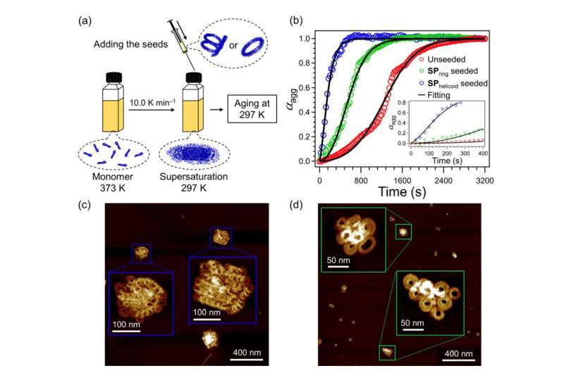
Depiction of the seeded supramolecular polymerization protocol (a) and the associated time-dependent changes (b). Supramolecular polymers obtained by open-ended and closed-ended seeds (c and d, respectively). Credit: Shiki Yagai from Chiba University
Supramolecular polymers are a new class of polymers that are currently being evaluated for material applications. These interesting compounds also play an important role in cellular activities in the body. “Supra,” as the name suggests, is attributed to some unique properties that go beyond those of conventional polymers. Unlike traditional polymers, which are held together by strong, irreversible covalent bonds, supramolecular polymers are held together by weaker, reversible hydrogen bonds.
Supramolecular polymers can reversibly assemble and disassemble, are highly versatile, and can be used for developing targeted drug delivery therapies, sensors to detect pollutants, diagnostic markers, energy storage devices, personal care products, and self-repairing and recyclable materials. While their excellent recyclability makes them wonderful candidate molecules for sustainable applications, there is one roadblock—researchers have yet to understand how to control their polymer growth.
However, there have been advancements in this aspect. Researchers are now able to build “unlikely” polymers by triggering their assembly with “seeds,” enabling control their polymer growth. There are two main mechanisms through which this seed-induced self-assembly occurs: primary nucleation or elongation, where the polymer grows from its end; and secondary nucleation, where new molecules join the polymer by sticking to its surface. The distinction between these processes is important because it enables researchers to better control and manipulate the growth of these unique polymers. Unfortunately, in most cases of seeded self-assembly, primary and secondary nucleation can be difficult to tell apart.
To tackle this issue, a group of researchers led by Professor Shiki Yagai from Chiba University aimed to compare and study the impact of these two processes while delineating the role of precisely controllable “seeded supramolecular polymerization.” Their goal was to figure out how different seed shapes affect the formation of new supramolecular polymers. Their findings are published in Chemical Communications.
Prof. Yagai tells us what motivated the team to pursue this topic of research: “Because of the difficulty in controlling polymerization, supramolecular polymers have not yet reached the point of practical application even though three decades have passed since their establishment as a concept.” He is convinced, however, that because of their versatility, further research in this area is likely to lead to widespread applications of these self-organizing polymers in our daily lives.
For their experiments, the researchers used two supramolecular polymers as “seeds.” While a closed-ended ring-shaped seed was used in a previous study, an open-ended, helicoidal seed was newly prepared. The researchers found that when the open-ended, helicoidal seed was used, it acted as a template for the target molecules to attach and grow longer. On the other hand, when the closed-ended ring-shaped seed was used, it did not elongate itself, but rather served as a surface where new molecules could attach and form clusters, like a platform for new structures.
This research shows that the type of seed used in self-assembling supramolecular polymers influences the way the molecules assemble, and the final shape of the formed structures. This opens up exciting possibilities for various applications, from self-repairing and more easily recyclable materials to more advanced drug delivery systems, sensing technologies, and energy storage devices.
Prof. Yagai states, “By understanding these assembly processes, we can design and develop the next generation of more precise and environmentally friendly polymers with tailored structures and properties. The practical application of supramolecular polymers will enable us to produce plastic materials with lower energy consumption and reduce the energy required for recycling.”
The ability to manipulate these versatile, self-assembling polymers at the molecular level offers great potential for addressing complex challenges and creating innovative, sustainable solutions in fields ranging from healthcare to environmental sustainability.
More information: Hiroki Itabashi et al, Distinct seed topologies enable comparison of elongation and secondary nucleation pathways in seeded supramolecular polymerization, Chemical Communications (2023). DOI: 10.1039/D3CC01587D
RNA, an essential biomolecule for life, has been used in environmental applications including monitoring microbial communities, developing pesticides, and quantifying the abundance of pathogenic viruses, such as SARS-CoV-2, in water and wastewater systems. Understanding how quickly RNA breaks down in given conditions is critical to harnessing the molecule in these and other emerging technologies.
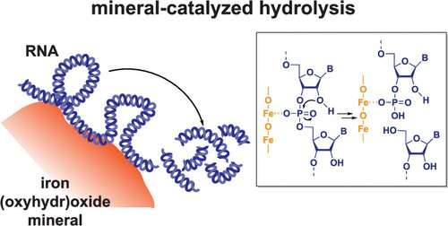
According to a new study by researchers working with Kimberly Parker, assistant professor of energy, environmental & chemical engineering in the McKelvey School of Engineering at Washington University in St. Louis, RNA can undergo rapid hydrolysis when adsorbed into iron oxide minerals. This discovery unveils a previously unknown abiotic pathway for RNA degradation and sheds light on biogeochemical processes and environmental system dynamics. The results were published May 22 in Environmental Science & Technology.
“This is the first abiotic process we’ve found that causes RNA degradation in the environment on timescales that can compete with biotic degradation,” Parker said. “Instead of depending on biological agents like enzymes or microbes to break down RNA molecules, we found that RNA degradation catalyzed by minerals happens relatively quickly regardless of the biological context. This could be an important limit on how long RNA persists in the environment.”
First author Ke Zhang conducted the research in Parker’s lab while earning a doctorate in environmental engineering at WashU in 2022. Zhang found that RNA undergoes rapid hydrolysis on the timescale of hours when adsorbed to iron oxide minerals such as goethite and hematite. This hydrolysis process is uniquely facilitated by the presence of iron in the minerals, which chemically accelerates the structural breakdown of the RNA molecule. This finding challenges scientists’ previous assumptions about the environmental factors affecting RNA degradation, particularly in iron-rich soils and sediments, which account for approximately 10% of global ice-free land.
“This process could provide an important limit on how long RNA hangs around in the environment, but there are certain conditions that can block this breakdown pathway,” Parker said. “While we measured the reaction timescales and determined the reaction products in this research, we need to develop more insights into the reaction mechanism in the future. Understanding the mechanisms as well as timescales of RNA degradation is crucial for accurately interpreting relative amounts of DNA versus RNA, studying viruses and pesticides, and even exploring the origin of life.”
More information: Ke Zhang et al, RNA Hydrolysis at Mineral–Water Interfaces, Environmental Science & Technology (2023). DOI: 10.1021/acs.est.3c01407
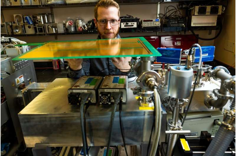
Adam Hollerbach with a SLIM device created at Pacific Northwest National Laboratory. Credit: Andrea Starr | Pacific Northwest National Laboratory
The universe is awash in billions of possible chemicals. But even with a bevy of high-tech instruments, scientists have determined the chemical structures of just a small fraction of those compounds, maybe 1%.
Scientists at the Department of Energy’s Pacific Northwest National Laboratory (PNNL) are taking aim at the other 99%, creating new ways to learn more about a vast sea of unknown compounds. There may be cures for disease, new approaches for tackling climate change, or new chemical or biological threats lurking in the chemical universe.
The work is part of an initiative known as m/q, or “m over q”—shorthand for mass divided by charge, which signifies one of the ways that scientists measure chemical properties in the world of mass spectrometry.
“Right now, we can take a sample from soil, where, depending on soil type, there may be thousands of chemical compounds in just a teaspoon’s worth,” said Thomas Metz, who leads the m/q Initiative. “And we don’t know what most of them are in terms of their chemical structures. We simply have no idea what’s in there.”
Scientists typically rely on reference libraries that contain information about thousands of molecules to identify substances. Researchers sort their samples from soil, the body, or elsewhere and compare what they have measured experimentally to what’s in the library. While that’s helpful, it limits scientists to only structurally identifying molecules that have been seen before—for example, through analysis of standard compounds purchased from chemical suppliers.
In the latest development, a team led by scientist Adam Hollerbach has combined two high-resolution instruments into one system to size up molecules in unprecedented detail. The results were published June 12 in the journal Analytical Chemistry.
Now, scientists can make several important measurements about chemical compounds in one experiment, gaining important information faster, more conveniently, and more accurately than before.
Hollerbach’s technique applies to ions—molecules that have either a positive or negative charge. That makes them easier to control and possible to detect using mass spectrometry.
Mass spectrometry: Tool of the ion whisperers
Like the people who study them, ions have many features that distinguish one from another. In people, weight, hair color, size, shape, eye color, and many other characteristics help us know who’s who. For ions, identifying characteristics include mass, shape, size, electric charge, and chemical composition. Those not only serve as identifiers but also as guides to the associated molecules’ behavior—clues to their potential to cure disease or sop up pollutants, for example.
That understanding should help the efforts of scores of scientists at PNNL who focus on understanding the effect of microbes on climate. Microbes play a key role in transforming elements like carbon into other forms that are important for the planet. Their impact on warming or cooling the planet is mighty. But scientists have much to learn.
“There may be millions of microbes in just a gram of soil, and we don’t know who most of them are or what they do. There’s a lot of discovery still to happen,” said Metz. “From the viewpoint of challenging science, it’s either a worst-case scenario or one of our greatest opportunities, depending on how you look at it.”
The m/q scientists are seizing the opportunity. Instead of framing their questions within the relatively small number of compounds that can be identified in conventional mass spectrometry measurements, they’re trying to leapfrog current limitations and create a whole new way of identifying what is unknown today. It’s a bit like when a new telescope is deployed and reveals several distinct stars where before, just one blurry hodgepodge of celestial bodies was visible.
The work is both experimental, putting molecules through their paces in the laboratory, and on computers, where scientists model what they are seeing and predict what they will likely see.
In the experiments described in the Analytical Chemistry paper, Hollerbach and colleagues made sensitive measurements of peptides and lipids. The experiments combined two instruments with similar names but that provide different details about ions. Both are used in mass spectrometry, a field whose history is interwoven with discoveries by PNNL scientists.
The first instrument is a mass spectrometer, which measures an ion’s mass, electric charge, and how the ion breaks apart. In this study, the team used an Orbitrap developed by Thermo-Fisher Scientific. Such instruments sort molecules of different masses well, but two molecules with the same mass are difficult to separate. Think of two people, each weighing 180 lbs.—one is tall and thin while the other is short and stocky. On a scale alone, they would be impossible to separate.
A SLIM approach: Ion mobility spectrometry brings hefty results
The second instrument is known as SLIM: structures for lossless ion manipulations. SLIM, created by PNNL scientist Richard D. Smith and colleagues, is an ion mobility spectrometer that measures an ion’s size and electric charge.
SLIM, which is about the size of a laptop and stands at just one-quarter of an inch thick, is a hothouse of molecular activity. Dozens of long, winding paths transform the small device into a 42-foot-long molecular racetrack, with ions that are controlled tightly by electric fields racing round and round an oval obstacle course.
The “obstacles” are other, known molecules such as helium or nitrogen molecules. As the ions under study race through the SLIM device, they navigate around or through the other molecules, tumbling and swerving much like a football running back runs through and around opposing blockers. The term “ion mobility spectrometry” truly captures the action.
By recording how long it takes for the ions to complete the course—how deftly they navigate the blocking ions—scientists learn all kinds of things about ions’ shape and size. That information, which isn’t available from a standard mass spec instrument, is combined with data about the ion’s mass, electric charge, and fragmentation pattern. Altogether, the data yields the ion’s collision cross section, its molecular formula, and its fragmentation pattern, properties that are central to understanding a molecule’s structure.
“Two different molecules can have the same number of atoms, and the same mass and charge, but they could have very different structures and activity. That’s where SLIM comes in to tell the difference,” said Hollerbach. “Just one small change can mean the difference between a molecule that is indicative of a disease and one that’s not.”
The key to Hollerbach’s experiment was getting the two different instruments to play nicely together. While both standard mass spectrometry and ion mobility spectrometry analyze ions, they work on different time scales. Ions make their journey through SLIM and arrive at the Orbitrap faster than they can be processed.
So Hollerbach drew on an old technique, deploying “dual-gated ion injection.” He added gates to control the intake of ions into the system and to control their arrival at the Orbitrap, choosing to send some of the ions from SLIM into oblivion to keep the flow at a manageable rate.
“Really, the questions we ask are very simple,” said Hollerbach. “What is this, and how much is there? But the techniques we use are complex.”
Other m/q scientists are working on additional ways to identify or exploit unknown molecules. Some are creating ways to use data like that from Hollerbach’s experiment to predict an ion’s structure automatically, so drug makers and other scientists would know exactly what they’re working with. Others are scouting out the millions of possibilities for forms of compounds such as fentanyl, sorting out what’s unlikely from what might show up on the street one day. Then they predict how those compounds would behave inside a mass spectrometer—creating a way to identify them if and when they do show up.
More information: Adam L. Hollerbach et al, A Dual-Gated Structures for Lossless Ion Manipulations-Ion Mobility Orbitrap Mass Spectrometry Platform for Combined Ultra-High-Resolution Molecular Analysis, Analytical Chemistry (2023). DOI: 10.1021/acs.analchem.3c00881
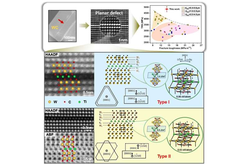
Composition, structure and crystallography of the new type planar defects. Credit: the authors
In collaboration with the Institute of Physics at the Chinese Academy of Sciences (IPCAS), researchers from Beijing University of Technology (BJUT) have discovered a new type of grain-interior planar defect in a ceramic phase in TiC doped cemented tungsten carbides.
These planar defects were found to be a result of the ordered distribution of heteroatoms on specific crystal planes of tungsten (W) and carbon (C). Importantly, these newly identified defects display distinct characteristics that set them apart from known planar defects, such as phase boundaries, grain boundaries, twin boundaries, stacking faults and complexions.
The work is published in Advanced Powder Materials, and involved detailed characterizations on the atomic scale for the composition, structure and crystallography of the new type of planar defects. In addition, comprehensive model calculations were conducted to assess the defects’ energy state and stability, providing further insights into their nature.
The research team, led by Professor Xiaoyan Song from BJUT, found that the occurrence of titanium (Ti) monolayer on the basal planes of WC was caused by the destabilization of (W,Ti)Cx complexions, which formed at the WC/Co interfaces by dissolution-precipitation processes during sintering of the powder mixture. The stable Ti monolayer may provide nucleation sites for the growth of WC crystal along the [0001]WC direction. This possibility was confirmed by model calculations.
“We further explored the possibility for the formation of such planar defects in WC grains by doping of V, Zr, Nb, Mo, and Hf through modeling,” explained Song. “We found that the Ti-monolayer induced planar defects had the highest stability in the WC grain interior, and also were much easier to form in the cemented carbides.”
The work highlights the significance of planar defects with high stability hindering the long-distance motion of stacking faults and dislocations within grains, and act as obstructions against propagation of the transgranular cracks. Thus, the risk of transgranular fracture, which is the dominant failure mode of the covalent crystals in ceramics and ceramic matrix composites, can be significantly reduced.
“We conclusively demonstrated that adjusting the density of planar defects allows for optimal mechanical performance, striking an exceptional balance between strength and fracture toughness in the materials,” added Song. “Our study paves the way for improving the mechanical properties of materials through the deliberate introduction and customization of heteroatomic monolayer-induced grain-interior planar defects.”
Furthermore, the methodology described in this article, using cemented tungsten carbides as a representative case, can be extended to other materials. By carefully selecting dopants and controlling sintering parameters, it becomes possible to fine-tune the density of planar defects to achieve desired properties in various ceramic systems.
More information: Xingwei Liu et al, Grain-interior planar defects induced by heteroatom monolayer, Advanced Powder Materials (2023). DOI: 10.1016/j.apmate.2023.100130
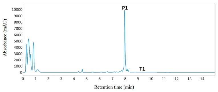
UPLC analysis of polymerase/endonuclease catalyzed P1 formation under optimized conditions. UPLC trace showing crude product P1 formed during the KOD polymerase (2 µM) and TnEndoV endonuclease (2 µM) catalyzed reaction using T1 (1 µM), dNTPs (4 mM each), in 20 mM Tris-HCl (pH 8), 20 mM MgCl2, 100 mM arginine-HCl (pH 8), 100 mM glutamate-KOH (pH 8), DTT (10 mM), formamide (10%), trehalose (0.2 M), 1,2-propanediol (1 M) and AcBSA (0.01 mg/ml) following 12 hours incubation at 70 °C. Product was generated following 330 cycles of template extension and product cleavage to afford 0.33 mM (equivalent to 1.9 g/L) of P1. Credit: Science (2023). DOI: 10.1126/science.add5892
A team of biochemists at the Manchester Institute of Biotechnology has developed an isothermal biocatalytic process that can be used to manufacture therapeutic oligonucleotides in large volumes. In their paper published in the journal Science, the group describes their process and possible medical applications.
Oligonucleotides (short base pairs of RNA or DNA molecules) have been used to treat several rare diseases. But the process of manufacturing them in large quantities is difficult for more general use. More recently, the development of therapies such as siRNA inclisiran to reduce cholesterol in the bloodstream has put pressure on biochemists to develop a less difficult process.
In this new effort, the team developed a one-pot manufacturing process. They note that it can be conducted in an aqueous solution and uses polymerase enzymes to extend template strands and an endonuclease enzyme to release the final product, allowing for the templates used in the process to be used repeatedly.
The process starts by adding nucleoside triphosphates to an aqueous solution, which drives a template-dependent synthetic reaction. Adding an extended template results in product cleavage, leading to the creation of endonuclease V, part of which can be removed for use as it is. The other part is then exposed to yet another template made using inosines, and the material that makes it through the final template consists of the desired oligonucleotides.
The research team suggests that in addition to making the manufacture of therapeutic oligonucleotides less expensive, it also makes it more scalable. To prove their claims, they used their new method to manufacture several well-known therapeutic oligonucleotides, such as pegaptanib, which has been used to treat macular degeneration.
They were able to produce approximately 2g per liter, but suggest the process should allow for amounts over 10g per liter—and at some point, as much as 100g per liter. They plan to continue fine tuning the process to manufacture more kinds of oligonucleotides. The team has partnered with Novartis, which already produces inclisiran, to further develop the scaling process.
More information: E. R. Moody et al, An enzyme cascade enables production of therapeutic oligonucleotides in a single operation, Science (2023). DOI: 10.1126/science.add5892

. DOI: 10.1016/j.carbon.2023.118215")
. DOI: 10.1021/acs.joc.3c00056")

. DOI: 10.1021/acsomega.3c01812")

 and Shuai Liu are lead co-authors on the study published in the Journal of the American Chemical Society. Credit: Gustavo Raskosky/Rice University")
 and the associated time-dependent changes (b). Supramolecular polymers obtained by open-ended and closed-ended seeds (c and d, respectively). Credit: Shiki Yagai from Chiba University")
. DOI: 10.1021/acs.est.3c01407")


 and TnEndoV endonuclease (2 µM) catalyzed reaction using T1 (1 µM), dNTPs (4 mM each), in 20 mM Tris-HCl (pH 8), 20 mM MgCl2, 100 mM arginine-HCl (pH 8), 100 mM glutamate-KOH (pH 8), DTT (10 mM), formamide (10%), trehalose (0.2 M), 1,2-propanediol (1 M) and AcBSA (0.01 mg/ml) following 12 hours incubation at 70 °C. Product was generated following 330 cycles of template extension and product cleavage to afford 0.33 mM (equivalent to 1.9 g/L) of P1. Credit: Science (2023). DOI: 10.1126/science.add5892")