How urea may have been the gateway to life

by ETH Zurich

origin of life
Credit: Unsplash/CC0 Public Domain

Researchers from ETH Zurich and the University of Geneva have developed a new method that allows them to observe chemical reactions taking place in liquids at extremely high temporal resolution. This means they can examine how molecules change within just a few femtoseconds—in other words, within a few quadrillionths of a second. The method is based on earlier work done by the same group of researchers led by Hans Jakob Wörner, Professor of Physical Chemistry at ETH Zurich. That work yielded similar results for reactions that take place in gas environments.

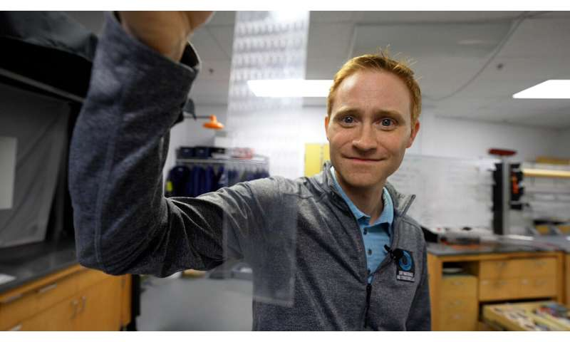
To expand their X-ray spectroscopy observations to liquids, the researchers had to design an apparatus capable of producing a liquid jet with a diameter of less than one micrometer in a vacuum. This was essential because if the jet were any wider, it would absorb some of the X-rays used to measure it. The work is published in the journal Nature.

Molecular pioneer in biochemistry

Using the new method, the researchers were able to gain insights into the processes that led to the emergence of life on Earth. Many scientists assume that urea played a pivotal role here. It is one of the simplest molecules containing both carbon and nitrogen. What’s more, it’s highly likely that urea was present even when the Earth was very young, something that was also suggested by a famous experiment done in the 1950s: American scientist Stanley Miller concocted a mixture of those gases believed to have made up the planet’s primordial atmosphere and exposed it to the conditions of a thunderstorm. This produced a series of molecules, one of which was urea.

According to current theories, the urea could have become enriched in warm puddles—commonly called primordial soup—on the then lifeless Earth. As the water in this soup evaporated, the concentration of urea increased. Through exposure to ionizing radiation such as cosmic rays, it’s possible that this concentrated urea produced malonic acid over multiple synthesis steps. In turn, this may have created the building blocks of RNA and DNA.

Why this exact reaction took place

Using their new method, the researchers from ETH Zurich and the University of Geneva investigated the first step in this long series of chemical reactions to find out how a concentrated urea solution behaves when exposed to ionizing radiation.

It’s important to know that the urea molecules in a concentrated urea solution group themselves into pairs, or what are known as dimers. As the researchers have now been able to show, ionizing radiation causes a hydrogen atom within each of these dimers to move from one urea molecule to the other. This turns one urea molecule into a protonated urea molecule, and the other into a urea radical. The latter is highly chemically reactive—so reactive, in fact, that it’s very likely to react with other molecules, thereby also forming malonic acid.

The researchers also managed to show that this transfer of a hydrogen atom happens extremely quickly, taking only around 150 femtoseconds, or 150 quadrillionths of a second. “That’s so fast that this reaction preempts all other reactions that might theoretically also take place,” Wörner says. “This explains why concentrated urea solutions produce urea radicals rather than hosting other reactions that would produce other molecules.”

Reactions in liquids are highly relevant

In the future, Wörner and his colleagues want to examine the next steps that lead to the formation of malonic acid. They hope this will help them to understand the origins of life on Earth.

As for their new method, it can also generally be used to examine the precise sequence of chemical reactions in liquids. “A whole host of important chemical reactions take place in liquids—not just all biochemical processes in the human body, but also a great many chemical syntheses relevant to industry,” Wörner says. “This is why it’s so important that we have now expanded the scope of X-ray spectroscopy at high temporal resolution to include reactions in liquids.”

The researchers from ETH Zurich and the University of Geneva were assisted in this work by colleagues from Deutsches Elektronen-Synchrotron DESY in Hamburg, who performed calculations required to interpret measurement data.

More information: Hans Jakob Wörner, Femtosecond Proton Transfer in Urea Solutions Probed by X-ray Spectroscopy, Nature (2023). DOI: 10.1038/s41586-023-06182-6www.nature.com/articles/s41586-023-06182-6

Journal information: Nature 

Provided by ETH Zurich 

The potential of generative AI to accelerate antiviral development and drug discovery

by Diamond Light Source

Breakthrough Research shows potential for generative AI to accelerate development of new antivirals and drug discovery
Surface representation of SARS CoV-2 Mpro protein with fragment hits from Diamond XChem platform bound in active site (Green). Credit: Diamond Light Source Ltd

In a new study, researchers from IBM, Oxford University and Diamond Light Source show that IBM’s AI Model, MoLFormer, can generate antiviral molecules for multiple target virus proteins, including SARS-CoV-2, that can accelerate the drug discovery process and bolster our response to future pandemics.

The results are laid out in a new paper published in Science Advances, and at the time of the paper’s submission, the antiviral properties of eleven molecules were successfully validated by Oxford researchers. This breakthrough has the potential to get drugs to people faster in the next crisis and bring treatments for urgent, life-threatening illnesses within reach.

Early in the pandemic, a group of computer scientists at IBM wanted to explore if generative AI could be used to design never-before seen molecules to block SARS-CoV-2, the virus that causes COVID-19. David Stuart, Head of the Division of Structural Biology in the Department of Clinical Medicine at the University of Oxford and Life Sciences Director at Diamond Light Source, the UK’s national synchrotron who is an authority on pathogens HIV, SARS, and Ebola, among other viruses explains he was initially skeptical. “The idea that you could take a protein sequence and, with AI, pluck out of thin air chemicals that would bind to a 3D site on the virus seemed very unlikely,” he said.

However, he and Martin Walsh also an expert structural biologist and Life Sciences Deputy director at Diamond joined up with the IBM team and over the course of three years, demonstrated that generative AI could, “pluck viable starting points for antivirals out of thin air,” in collaboration with Enamine Ltd., a chemical supplier in Ukraine, and other researchers at Oxford.

Because the generative model was also a foundation model, pre-trained on massive amounts of raw data, it was versatile enough to create new inhibitors for multiple protein targets without extra training or any knowledge of its 3D structure.

The Stuart and Walsh groups had commenced working on two essential SARS-CoV-2 proteins, namely the spike protein and the main protease. Using these targets, the team hit on four potential COVID-19 antivirals in a fraction of the time it would have taken using conventional methods. The work then exploited Diamond’s high-throughput macromolecular crystallography beamlines to visualize how a subset of the AI generated compounds bound to the main protease.

Their work is showcased in their new paper in Science Advances and IBM has released a web-based interface for interacting with the model and chemical foundation models like it in IBM Cloud.

The team stated that the validated molecules have many more hurdles to clear, including clinical trials, before companies could potentially turn them into drugs. But even if the AI-generated “hits” never materialize into actual drugs, the work provides confirmation that generative AI has an important role to play in the future of drug development, especially in a time of crisis.

“It took time to develop and validate these methods, but now that we have a working pipeline in place, we can generate results much faster,” said study co-senior author, Payel Das, a researcher at IBM Research. “When the next virus emerges, generative AI could be pivotal in the search for new treatments.”

“Generating initial compounds that bind with high affinity to a drug target of interest accelerates the structure-based drug discovery pipeline and underpins our efforts to be better prepared for future pandemics,” said, Martin Walsh, who was co-senior author at Diamond

The researchers built their model, Controlled Generation of Molecules (or CogMol), on a generative AI architecture known as variational autoencoders, or VAEs. VAEs encode raw data into a compressed representation, and then decode, or translate, it back into a statistical variation on the original sample. Their model was trained on a large dataset of molecules represented as strings of text, along with general information about proteins and their binding properties. But they deliberately left out information about SARS-CoV-2’s 3D structure or molecules known to bind to it. Their goal was to give their generative foundation model a broad base of knowledge so that it could be more easily deployed for molecular design tasks it has never seen before.

Their goal was to find drug-like molecules that would bind with two COVID protein targets: the spike, which transmits the virus to the host cell, and the main protease, which helps to spread it. Though the 3D structures of both proteins had been discovered by that time, the IBM researchers chose to use only their amino acid sequences, derived from their DNA. By limiting themselves in this way, they hoped that the model could learn to generate molecules without knowing the shape of their target.

The researchers input only the amino acid sequence for each protein target into CogMol, which generated 875,000 candidate molecules in three days. To narrow the pool, the researchers ran the candidates through a retrosynthesis platform, IBM RXN for Chemistry, to understand what ingredients would be needed to synthesize the compounds. Based on the platform’s predicted recipes, they selected 100 molecules for each target. Chemists at Enamine further pared the list to four molecules for each target, selecting those deemed easiest to manufacture.

After synthesizing the eight novel molecules, Enamine shipped them to Oxford for testing their ability to disrupt the functions of the two protein targets in the labs of Prof Chris Schofield and PRof Gavin Screaton. . The intense X-ray beam generated from Diamond which are 10 billion times brighter than the sun were used to visualize how the compounds interacted with proteins to inactivate their function. The novel compounds were further tested in target inhibition and live virus neutralization tests. Two of the validated antivirals target the main protease; the other two not only targeted the spike protein but proved capable of neutralizing all six major COVID variants. “You get a map that shows exactly where things bind, and bang! you’ve got a confirmation,” said Stuart.

Breakthrough Research shows potential for generative AI to accelerate development of new antivirals and drug discovery
Diamond Light Source, UK’s national synchrotron aerial View. Credit: Diamond Light Source Ltd 2021

CogMol is one of several chemical foundation models that IBM has since developed. The largest, MoLFormer-XL, was trained on a database of more than 1.1 billion molecules and is currently being used by Moderna to design mRNA medicines. “We created valid starting points for accelerated development of antivirals using a generative foundation model that knew relatively little about its protein targets,” said the study’s co-senior author, Jason Crain, a researcher at IBM Research and professor at Oxford. “I’m hopeful that these methods will allow us to create antivirals and other urgently needed compounds much faster and more inexpensively in the future.”

Though the researchers focused on validating antivirals for COVID, they argue that these methods can be extended to existing viruses that continue to mutate, like the flu, or viruses that have yet to surface. “If you want to be prepared for the next pandemic, you want drugs that act on different sites of the protein,” concluded Stuart. “It becomes much harder for the virus to escape.”

More information: Vijil Chenthamarakshan et al, Accelerating drug target inhibitor discovery with a deep generative foundation model, Science Advances (2023). DOI: 10.1126/sciadv.adg7865

Journal information: Science Advances 

Provided by Diamond Light Source 

New light-responsive carriers for intracellular substance delivery

by Okayama University

Towards new light-responsive carriers for intracellular substance delivery
LiDLs demonstrate high stability in extracellular compartments and low stability in intracellular compartments, effectively resolving the challenge faced by conventional drug delivery systems. As a result, they are promising photoswitchable carriers for intracellular substance delivery, find researchers from Japan in a new study. Credit: Yuki Sudo from Okayama University, Japan

Artificial lipid bilayer vesicle liposomes, also called proteoliposomes, are specialized systems capable of incorporating various molecules, such as chemicals and drugs. Their unique properties make them ideal carriers for delivering substances inside cells. However, they must possess the dual characteristics of high stability in extracellular environments and low stability in intracellular environments.

Several techniques have been developed to regulate the stability of liposomes in a condition-dependent manner, with pH-sensitive liposomes being widely employed. A standard measure of acidity or basicity, the pH scale ranges from 1 to 14, with 7 standing for “neutral,” like water, a pH below 7 indicating acidity and that above 7 indicating basicity. Interestingly, pH-sensitive liposomes can be triggered to release their contents when exposed to an acidic pH below 5.5.

In a new study, a team of researchers, led by Professor Yuki Sudo from Okayama University, Japan, have developed a precise method of releasing contents from pH-sensitive liposomes using light. “The study presents a novel nanomaterial called light-induced disruptive liposome (LiDL) and applies it to light-controlled intracellular substance delivery,” explains Prof. Sudo. Their work, co-authored by Mr. Taichi Tsuneishi of Okayama University and Prof. Yuma Yamada of Hokkaido University in Japan, was published in the journal Chemical Communications .

The researchers utilized a protein called Rubricoccus marinus Xenorhodopsin (RmXeR), derived from a marine bacterium, to initiate acidification inside liposomes using light. To study the functionality of RmXeR within liposomes without interference from pH-dependent properties, they first developed pH-insensitive proteoliposomes using the lipid hydration method, combining phosphatidylcholine from egg yolk with cholesterol. Then, purified RmXeR was incorporated via the dilution method. The researchers estimated that the pH inside the thus obtained liposomes changed from 7.0 to 4.8 upon exposure to green light, rendering them suitable for light-induced disruption.

Subsequently, the team developed pH-sensitive proteoliposomes based on 1,2-dioleoyl-sn-glycero-3-phosphoethanolamine and cholesteryl hemisuccinate. Interestingly, before introducing RmXeR within the liposomes, the researchers incorporated a water-soluble fluorescent dye called calcein as a testing substance to evaluate their release capability.

By monitoring the fluorescence of calcein, they observed that its release from the liposomes occurred in a light-dependent manner, indicating that the liposomes effectively released substances when triggered by light. These pH-sensitive proteoliposomes, or LiDLs, displayed exceptional stability in the absence of light, maintaining their physiochemical properties when exposed to a temperature of 4°C for two weeks.

In the next phase of the study, the researchers introduced LiDL into mammalian HeLa cells to evaluate their effectiveness as carriers for delivering substances inside the cells. They monitored the cellular uptake for LiDLs, finding that the presence of LiDL, regardless of the inclusion of RmXeR, enhanced the uptake of liposomes by HeLa cells. This demonstrated the potential of LiDL as a carrier for intracellular substance delivery. To further improve the intracellular delivery efficiency, the researchers introduced a chemical called stearyl octaarginine into LiDL. This enhanced version, called LiDL-R8, exhibited significantly better performance compared to LiDL.

These findings highlight the utility of LiDL for the intracellular delivery of chemicals as well as biomolecules. “With LiDL, drug delivery can be controlled by light, potentially leading to advances in various therapies,” emphasizes Prof. Sudo. Going forward, the team aims to enhance the content release efficiency by thoroughly characterizing the structure of the liposomes and optimizing the experimental conditions.

In effect, LiDLs hold great promise as optically controllable carriers for improved drug delivery. These liposomes offer enhanced efficacy without causing side effects, showcasing exceptional extracellular stability and controllable intracellular instability. “Our study will contribute towards ensuring healthy lives and promoting well-being for people of all ages, which ties in with one of the Sustainable Development Goal (SDG3) of the United Nations,” concludes Prof. Sudo.

More information: Taichi Tsuneishi et al, Development of light-induced disruptive liposomes (LiDL) as a photoswitchable carrier for intracellular substance delivery, Chemical Communications (2023). DOI: 10.1039/D3CC02056H

Journal information: Chemical Communications 

Provided by Okayama University 

How do ceramic particles strengthen metallic copper?

by International Journal of Extreme Manufacturing

How do ceramic particles strengthen metallic copper?
Various preparation techniques and characteristics for advanced powder metallurgy. (a) 
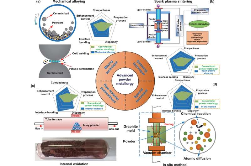
Mechanical alloying, (b) spark plasma sintering. Reproduced from [29]. CC BY 3.0. (c) Internal oxidation. Reprinted from [30], Copyright (2019), with permission from Elsevier.(d) In-situ processing. Credit: International Journal of Extreme Manufacturing (2023). DOI: 10.1088/2631-7990/acdb0b

Publishing in the International Journal of Extreme Manufacturing, scientists from Jilin University and Edith Cowan University comprehensively reviewed the latest preparation techniques of copper matrix composites and the effect of ceramic particles on the mechanical properties, thermal conductivity and thermal expansion behavior of the composites. Four main aspects of particle characterization were included: particle content, particle size, particle morphology and interfacial bonding of particles to copper matrix.

Team leader, Feng Qiu, said, “By reviewing the preparation techniques and effect mechanisms of particle reinforced copper matrix composites, it is hoped that this will serve as a basis for more precise design and manipulation of composite microstructure to meet the growing demand for copper matrix composites in a wide range of application fields.”

So far, endeavors have been focusing on how to choose suitable ceramic components and fully exert the strengthening effect of ceramic particles in the copper matrix. Currently, the preparation of copper matrix composites by powder metallurgy is the most mature technique, but it still faces many unresolved process drawbacks.

Mechanical alloying and spark plasma sintering have a more prominent contribution to the improvement of grain refinement and densification of composite, as well as the dispersion of particles. While the internal oxidation and in-situ method could greatly enhance the interfacial bonding between the ceramic phase and the metal matrix. Further combinations of these advanced preparation techniques and the full utilization of each technical advantage remain to be explored in more detail.

The co-leading author, Prof. Hongyu Yang, added that, “practical challenges in manipulating particle characteristics reinforce the value of the preparation and mechanistic exploration, which contribute to further optimize the physical and chemical performance of composites.”

“Currently, the manipulation of ceramic particles in most copper matrix composites is mainly focused on particle content and size, while particle distribution, morphology, and interfacial bonding still need to be optimized, especially for the exploration of in-situ synthesis techniques.”

First author, Dr. Yifan Yan said, “Regarding the study of the effect mechanism of ceramic particle reinforced copper matrix composites, while experimental studies and mathematical models can reflect the influence of particle characteristics on material properties, finite element simulations provide a more intuitive perspective for predicting and studying the mechanism of intensifying action. However, optimal design of composites by computational simulation techniques in the definition of the constitutive model and the precise reconfiguration of the composite are largely unexplored.”

Prof. Laichang Zhang, an expert in advanced materials and also the co-leading author of the article, said, “Ongoing research in the field of ceramic reinforced copper matrix composites has yielded promising results in recent studies. Composites mixed with high-performance carbon nanotubes, carbon fibers, and advanced MAX-phase ceramic materials have demonstrated favorable comprehensive performance. However, there is limited information available on the interaction and distribution of strengthening phases.”

“Selecting and manipulating the strengthening phases in a rational manner could meet the performance requirements of copper matrix composites in various applications, but presents significant challenges in the design and preparation of composites. More complex material configuration designs, such as network structures and gradient structures, offer new opportunities to prepare anisotropic functional materials or speciality composites.”

More information: Yi-Fan Yan et al, Ceramic particles reinforced copper matrix composites manufactured by advanced powder metallurgy: preparation, performance, and mechanisms, International Journal of Extreme Manufacturing (2023). DOI: 10.1088/2631-7990/acdb0b

Provided by International Journal of Extreme Manufacturing

‘Designer’ tubulins help crack the tubulin code

by Ecole Polytechnique Federale de Lausanne

Cracking the tubulin code
An illustration of tubulin handled by a metaphorical pair of tweezers. Credit: Beat Fierz (EPFL)

Tubulin is a protein that plays a crucial role in the structure and function of cells. It is the main component of microtubules, which are long, hollow fibers that provide structural support, help the cell divide, give it its shape, and act as tracks for moving molecular cargo around inside the cell.

There are two types of tubulin: alpha-tubulin and beta-tubulin. Together, they form dimeric (two-part) building blocks, spontaneously assembling into microtubules that undergo further continuous cycles of assembly and disassembly.

To fine-tune microtubules, the dimers undergo various post-translational modifications (PTMs), which are chemical modifications that occur after they are synthesized, and can affect their structure, activity, and interactions with other molecules.

Two important PTMs take place on the unstructured tail of alpha-tubulin: Polyglutamylation, which adds chains of glutamate amino acids, and detyrosination, which removes the final tyrosine amino acid. These PTMs, among others, are found together in stable microtubules, e.g. in neurons.

Combinations of PTMs form what scientists refer to as a “tubulin code,” which is connected to specific functions of microtubules. Tubulin PTMs are critical for the proper functioning of microtubules.

Dysregulation of PTMs have been linked to various diseases, including cancer, neurodegeneration, and developmental disorders. Therefore, understanding the importance of tubulin PTMs is crucial for advancing our knowledge of these diseases and developing potential therapies. The problem is that the mechanisms that govern such PTM patterns are not well understood, mostly because we don’t have the tools to dissect the function and regulation of tubulin PTMs.

Cracking the tubulin code

Scientists at EPFL and the University of Geneva (UNIGE) have now developed a chemical method to engineer fully functional tubulin carrying precise combinations of post-translational modifications (PTMs). The study was led by Professors Beat Fierz (EPFL) and Assistant Professor Charlotte Aumeier (UNIGE), in collaboration with the labs of Pierre Gönczy (EPFL) and Carsten Janke (Institute Curie), and provides insight into how specific PTMs regulate the function of tubulin in cells. Their findings have been published in Nature Chemistry.

The method uses chemo-enzymatic protein splicing to attach synthetic alpha-tubulin tails that were modified with varying degrees of polyglutamate to human tubulin molecules. Using these “designer” tubulins allowed the researchers for the first time to assemble homogenously modified microtubules.

The researchers also found that polyglutamylation of alpha-tubulin facilitated its detyrosination by enhancing the activity of the protein complex vasohibin/SVBP, the key enzyme responsible for this modification. The team confirmed their findings by changing the levels of polyglutamate in living cells and observing the effects on tyrosine removal.

The study presents a novel approach to designing tubulins with specific PTMs and uncovers a new interplay between two key regulatory systems that control the function of tubulin: polyglutamylation and detyrosination.

The new method of producing tubulins with defined PTMs can advance our understanding of their molecular function, and provide insights into how dysregulation of these PTMs leads to diseases.

Based on this work, the labs of Fierz and Aumeier, together with Jens Stein at the University of Fribourg and Michael Sixt at ISTA Vienna will next investigate how tubulin PTMs control the cytoskeleton in migrating immune cells.

More information: Eduard Ebberink et al. Tubulin engineering by semi-synthesis reveals that polyglutamylation directs detyrosination, Nature Chemistry (2023). DOI: 10.1038/s41557-023-01228-8www.nature.com/articles/s41557-023-01228-8

Journal information: Nature Chemistry 

Provided by Ecole Polytechnique Federale de Lausanne 

New criteria to determine whether shear bands are beneficial or harmful to crystalline materials

by Jason Daley, University of Wisconsin-Madison

With sheer determination, researchers can make tough materials that bend without breaking
Professor Izabela Szlufarska (standing)
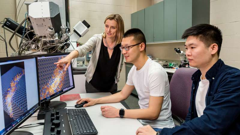
, and graduate students Xuanxin Hu(middle) and Nuohao Liu (right) have developed new criteria for determining whether shearbands are beneficial or harmful to certain crystalline materials. Credit: University of Wisconsin–Madison

Shear band formation is not typically a good sign in a material—the bands often appear before a material fractures or fails. But materials science and engineering researchers at the University of Wisconsin–Madison have found that shear bands aren’t always a negative; under the right conditions, they can improve the ductility, or the plasticity, of a material.

Led by Izabela Szlufarska, a professor of materials science and engineering at UW–Madison, the researchers published details of their work in the journal Nature Materials.

Using a combination of experimental characterization and simulations, the team identified potential strategies for encouraging shear bands. This could lead to new ways of increasing the toughness of a wide array of materials.

“In a previous paper, we demonstrated that shear bands in a material called samarium cobalt could actually be beneficial,” says Szlufarska. “That led to the questions, “When do shear bands form?” and “When do they support plasticity versus fracture? When do you want to avoid them and when do you want to promote them?'”

In materials with crystalline structures, like metals and ceramics, plasticity is determined by small structural irregularities within the crystal lattice called dislocations. These dislocations can move through the lattice and provide some give, which allows materials like metals to bend without ripping apart the bonds in their rigid structure. However, the more these dislocations are locked into place, either through hardening techniques or natural structural variations, the more brittle a material becomes.

That’s why Szlufarska and her team were surprised to find that in samarium cobalt, a brittle intermetallic material used to make strong magnets, amorphous, or unstructured, shear bands increased the material’s plasticity and were not a sign of failure. Instead, these areas acted as a lubricant, allowing planes of atoms to slide over one another without creating a fracture.

The team hypothesized that these types of beneficial shear bands can form in materials that easily transition between crystalline and amorphous phases. To test this, they looked at aluminum samarium, a glassy material studied extensively by Szlufarska and her colleagues in the Materials Research Science and Education Center at UW–Madison. Using atomic-level simulations, Szlufarska’s group predicted that the crystalline form of this material should also form shear bands under stress. They not only confirmed the finding in the lab, but also varied the atomic composition of the aluminum samarium, making versions where shear bands led either to fracture or to plasticity.

That understanding led the team to propose criteria for screening new materials that might exhibit similar properties and for identifying when shear bands are beneficial. The hope is that these parameters will make it possible to search databases to identify materials that could benefit from doping or engineering to promote shear band formation.

“Increasing toughness, or the amount of energy or stress a material can take before it breaks, by two or three or four times could be really powerful,” says Szlufarska. “In this paper, we extended our finding to a new class of materials. But this is not the end.”

The team intends to test traditional structural materials like oxides, carbides and borides to determine how they can be optimized.

“We understand so much more about how this happens and what to look for,” says Szlufarska. “I think we have identified the key to the design of these materials, and we want to now take them to other classes.”

Szlufarska has also published related research in the journal npj 2D Materials and Applications

More information: Jun Young Kim et al, Experimental and theoretical studies of native deep-level defects in transition metal dichalcogenides, npj 2D Materials and Applications (2022). DOI: 10.1038/s41699-022-00350-4

Journal information: Nature Materials 

Provided by University of Wisconsin-Madison 

Scientists discover that water molecules define the materials around us

Columbia scientists discover that water molecules define the materials around us
Spirit Island, Jasper National Park, Canada. “When we take a walk in the woods, we think of the trees and plants around us as typical solids,” professor Ozgur Sahin said. “This research shows that we should really think of those trees and plants as towers of water holding sugars and proteins in place.” Credit: Terry Ott

For decades, the fields of physics and chemistry have maintained that the atoms and molecules that make up the natural world define the character of solid matter. Salt crystals get their crystalline quality from the ionic bond between sodium and chloride ions, metals like iron or copper get their strength from the metallic bonds between iron or copper atoms, and rubbers get their stretchiness from the flexible bonds within polymers that constitute the rubber. The same principle applies for materials like fungi, bacteria, and wood.

Or so the story goes.

A new paper published in Nature upends that paradigm, and argues that the character of many biological materials is actually created by the water that permeates these materials. Water gives rise to a solid and goes on to define the properties of that solid, all the while maintaining its liquid characteristics. In their paper, the authors group these and other materials into a new class of matter that they call “hydration solids,” which they say “acquire their structural rigidity, the defining characteristic of the solid state, from the fluid permeating their pores.” The new understanding of biological matter can help answer questions that have dogged scientists for years.

“I think this is a really special moment in science,” Ozgur Sahin, a professor of Biological Sciences and Physics and one of the paper’s authors, said. “It’s unifying something incredibly diverse and complex with a simple explanation. It’s a big surprise, an intellectual delight.”

Steven G. Harrellson, who recently completed doctoral studies in Columbia’s physics department, and is an author on the study, used the metaphor of a building to describe the team’s finding: “If you think of biological materials like a skyscraper, the molecular building blocks are the steel frames that hold them up, and water in between the molecular building blocks is the air inside the steel frames. We discovered that some skyscrapers aren’t supported by their steel frames, but by the air within those frames.”

“This idea may seem hard to believe, but it resolves mysteries and helps predict the existence of exciting phenomena in materials,” Sahin added.

When water is in its liquid form, its molecules strike a fine balance between order and disorder. But when the molecules that form biological materials combine with water, they tip the balance toward order: Water wants to return to its original state. As a result, the water molecules push the biological matter’s molecules away. That pushing force, called the hydration force, was identified in the 1970s, but its impact on biological matter was thought to be limited. This new paper’s argument that the hydration force is what defines the character of biological matter almost entirely, including how soft or hard it is, thus comes as a surprise.

We have long known that biological materials absorb ambient moisture. Think, for example, of a wooden door, that expands during a humid spell. This research, however, shows that that ambient water is much more central to wood, fungi, plants, and other natural materials’ character than we had ever known.

The team found that bringing water to the front and center allowed them to describe the characteristics that familiar organic materials display with very simple math. Previous models of how water interacts with organic matter have required advanced computer simulations to predict the properties of the material. The simplicity of the formulas that the team found can predict these properties suggests that they’re onto something.

To take one example, the team found that the simple equation E=Al/λ neatly describes how a material’s elasticity changes based on factors including humidity, temperature, and molecule size. (E in this equation refers to the elasticity of a material; A is a factor that depends on the temperature and humidity of the environment; l is the approximate size of biological molecules and λ is the distance over which hydration forces lose their strength).

“The more we worked on this project, the simpler the answers became,” Harrellson said, adding that the experience “is very rare in science.”

The new findings emerged from Professor Sahin’s ongoing research into the strange behavior of spores, dormant bacterial cells. For years, Sahin and his students have studied spores to understand why they expand forcefully when water is added to them and contract when water is removed. Several years ago, Sahin and colleagues garnered media coverage for harnessing that capability to create small engine-like contraptions powered by spores.

Around 2012, Sahin decided to take a step back to ask why the spores behave the way they do. He was joined by researchers Michael S. DeLay and Xi Chen, authors on the new paper, who were then members of his lab. Their experiments did not provide a resolution to the mysterious behavior of spores. “We ended up with more mysteries than when we started,” Sahin remembers. They were stuck, but the mysteries they encountered were hinting that there was something worth pursuing.

After years of pondering potential explanations, it occurred to Sahin that the mysteries the team continually encountered could be explained if the hydration force governed the way that water moved in spores.

“When we initially tackled the project, it seemed impossibly complicated. We were trying to explain several different effects, each with their own unsatisfying formula. Once we started using hydration forces, every one of the old formulas could be stripped away. When only hydration forces were left, it felt like our feet finally hit the ground. It was amazing, and a huge relief; things made sense,” he said.

The paper’s findings apply to huge amounts of the world around us: Hygroscopic biological materials—that is, biological materials that allow water in and out of them–potentially make up anywhere from 50% to 90% of the living world around us, including all of the world’s wood, but also other familiar materials like bamboo, cotton, pine cones, wool, hair, fingernails, pollen grains in plants, the outer skin of animals, and bacterial and fungal spores that help these organisms survive and reproduce.

The term coined in the paper, “hydration solids,” applies to any natural material that’s responsive to the ambient humidity around it. With the equations that the team identified, they and other researchers can now predict materials’ mechanical properties from basic physics principles. So far that was true mainly of gases, thanks to the well-known general gas equation, which has been known to scientists since the 19th century.

“When we take a walk in the woods, we think of the trees and plants around us as typical solids. This research shows that we should really think of those trees and plants as towers of water holding sugars and proteins in place,” Sahin said, “It’s really water’s world.”

More information: Ozgur Sahin, Hydration solids, Nature (2023). DOI: 10.1038/s41586-023-06144-ywww.nature.com/articles/s41586-023-06144-y

Journal information: Nature 

Provided by Columbia University 

Tailoring fluorine-rich solid electrolyte interphase to boost long cycling stability of lithium metal batteries

Tailoring fluorine-rich solid electrolyte interphase to boost high efficiency and long cycling stability of lithium metal batter
Schematics illustrating the role of DFEC in improving solid electrolyte interphase (SEI) 
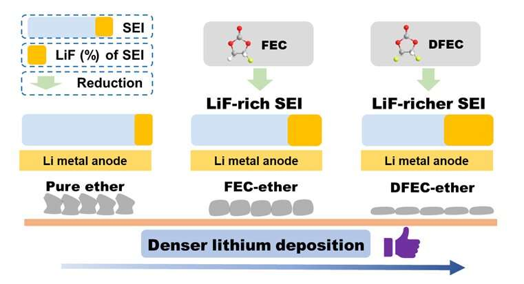
and affection on Li deposition in ether electrolyte. Credit: Science China Press

In a study published in the journal Science China Chemistry, fluorinated cyclic carbonate (DFEC) was introduced into ether electrolyte as a SEI-forming additive. The modified electrolyte could improve the interface of Li metal anode and achieve high efficiency and long cycling stability of LMBs.

LMBs are regarded as the most promising next-generation battery system due to the high specific capacity (3860 mAh g−1) and low electrode potential (-3.04 V vs. SHE) of the Li metal anode.

However, there are many limiting factors which limit the development of LMBs—such as a side reaction between Li anode and electrolyte, Li dendrite growth and a serious volume effect of Li anode, etc.—which lead to low coulombic efficiency (CE) and poor cycle life. Stable solid electrolyte interphase (SEI) is the key to achieve high efficiency and long cycling stability of LMBs.

Adjusting SEI through electrolyte optimization is regard as a low-cost and efficient way to improve Li metal anode interface. So, it is critical to design an electrolyte formulation which can form a stable SEI, the key is the choice of solvents and film-forming additive.

Recently, Prof. Renjie Chen and Prof. Ji Qian proposed an ether-ester mixed electrolyte in which trans-difluoroethylene carbonate (DFEC) was introduced into the ether electrolyte as a film-forming additive. Firstly, ether electrolyte has good anti-reduction stability with Li metal. Secondly, due to the lower LUMO level of DFEC, it can be preferentially reduced during the initial cycle, forming LiF-rich SEI on the Li metal anode.

LiF-rich SEI can inhibit the growth of lithium dendrite, alleviate side reactions, and induce dense lithium deposition. Thanks to the above advantages, the LMBs using modified electrolyte show high efficiency and stable cycling performance. The first author of this paper is Tianyang Xue, a graduate student at Beijing Institute of Technology, and the corresponding authors are Prof. Renjie Chen, Prof. Ji Qian, and Prof. Xingming Guo.

A few implications thus emerge for designing an electrolyte to boost high efficiency and long cycling stability of LMBs. This work explores the interphase chemistry of LMBs, and provides important insights for further study on the novel electrolyte system for LMBs.

More information: Tianyang Xue et al, Tailoring fluorine-rich solid electrolyte interphase to boost high efficiency and long cycling stability of lithium metal batteries, Science China Chemistry (2023). DOI: 10.1007/s11426-022-1623-2

Provided by Science China Press 

Mass spectrometry uncovers actions of protein ‘glues’

Mass spectrometry uncovers actions of protein 'glues'
(A) 
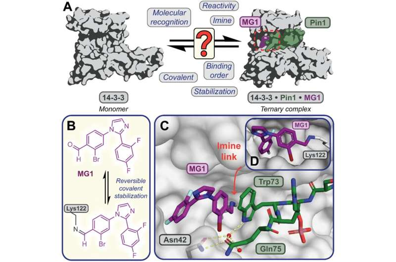
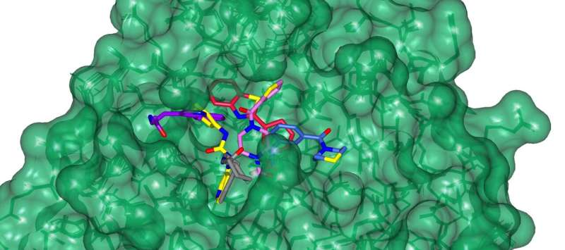
Illustrates the limited understanding of MG1 induced complex stabilization. (B) Schematic representation of MG1 ligation. (C) Enlarged view of the 14-3-3/Pin1/MG1 interface . (D) MG1 covalently bound to Lys122 of 14-3-3 by aldimine bonding. Credit: Chemical Science (2023). DOI: 10.1039/D3SC01732J

A screening technique commonly used in drug discovery can yield important details about the actions of molecular ‘glues’ in protein interactions.

Molecular glues are emerging as powerful therapeutic tools that can stick proteins together in the body. The interactions between proteins underpin all biological cell functions, including those of disease, and so interventions that can control protein-protein interactions have significant potential for disrupting the progress of various diseases.

While in many cases, drugs are required to interrupt the processes that connect proteins together, there are also occasions when the intervention is needed to restore an interaction, or to make it function correctly.

Researchers at the University of Birmingham, together with partners at the University of Leicester and the Eindhoven University of Technology, have devised a way of using mass spectrometry to analyze candidate glues for these processes and assess their relative strengths.

Dr. Aneika Leney, of the School of Biosciences at the University of Birmingham, explained, “Often when we are designing new drugs, it is to stop harmful protein interactions in the body, such as those that lead to tumor cell growth in cancers. Sometimes, however, the disease is caused by protein interactions falling apart and in these cases finding the right glue to hold them together could be extremely beneficial.”

In a new study, published in Chemical Science, the research team focused on one particular molecular glue, called MG1. Using the mass spectrometry method, they were able to disentangle the different mechanisms through which the glue bound to the proteins and stabilized the protein interaction. The MS method also allowed the researchers to elucidate the relative time taken by the different processes involved.

Dr. Peter Cossar, from the Department of Biomedical Engineering at Eindhoven University of Technology further explained, “Understanding how molecular glues stick proteins together enables scientist to better design and build the next generation of molecular glue drugs. Mass Spectrometry provides a tool to do so, by providing high fidelity information on how these unique molecules behave in real time.”

The team expect that the research will provide a robust framework for testing a wide range of molecular glues, offering a significant advance in drug discovery understanding in this area.

More information: Carlo J. A. Verhoef et al, Tracking the mechanism of covalent molecular glue stabilization using native mass spectrometry, Chemical Science (2023). DOI: 10.1039/D3SC01732J

Journal information: Chemical Science 

Provided by University of Birmingham 

Stronger tape engineered through the ancient Japanese art of cutting paper, kirigami

by Virginia Tech

Stronger tape engineered through the art of cutting
Associate Professor Michael Bartlett pulls enhanced tape developed in his lab at Virginia Tech. Credit: Alex Parrish for Virginia Tech.

Adhesive tape fulfills many purposes, from quickly fixing household appliances to ensuring a reliable seal on a mailed package. When using tape with a strong bond, removing it may only be possible by scraping and prying at the tape’s corners, hoping desperately that surface pieces don’t tear away with the tape.

But what if you could make adhesives both strong and easily removable? This seemingly paradoxical combination of properties could dramatically change applications in robotic grasping, wearables for health monitoring, and manufacturing for assembly and recycling.

Developing such adhesives may not by that far off through the latest research conducted by the team of Michael Bartlett, assistant professor in the Department of Mechanical Engineering at Virginia Tech, and published in Nature Materials on June 22.

The physics of stickiness

Adhesive tapes were first developed in the 1920s to meet a need for automobile painters who wanted better options for painting two colors on car bodies. Since the first masking tape was put into use, many other variations have been created. Factories have rolled out invisible tape for wrapping presents, electrical tape for covering wires, and duct tape for more uses than it was ever intended to fill.

Associate Professor Michael Bartlett holds altered tape developed in his lab at Virginia Tech. Credit: Alex Parrish for Virginia Tech.

Normally, when tapes are peeled off, they separate in a straight line along the length of the strip until the tape is completely removed. Strong adhesives are made more difficult to peel, while reusable adhesives promote the strength-limiting separation.

Bartlett’s team theorized that if the separation path were controlled, then perhaps adhesives could be made both strong and removable. They tapped into the methods of a 2,000-year-old Japanese art form to determine how to do it.

More information: Dohgyu Hwang et al, Metamaterial adhesives for programmable adhesion through reverse crack propagation, Nature Materials (2023). DOI: 10.1038/s41563-023-01577-2 , dx.doi.org/10.1038/s41563-023-01577-2

Journal information: Nature Materials 

Provided by Virginia Tech