Team develops novel tridimensional anticancer agents to fight against drug-resistant cancer cells

Novel tridimensional anticancer agents developed to fight against drug-resistant cancer cells
The half maximal inhibitory concentration (IC50 value) 
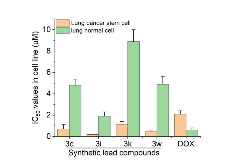
of synthetic lead compounds and clinical drug doxorubicin (DOX) in lung cancer stem cells and normal cells after incubation for 72 hours. Credit: Dr Zhu Guangyu’s Research Group / City University of Hong Kong

A research team co-led by chemists from City University of Hong Kong (CityU) recently discovered novel, highly effective anticancer agents with tridimensional structures, which have high anticancer activity, low toxicity and the ability to overcome drug resistance in cancer cells. The findings help provide a new direction for anticancer drug development.

Cancer has long been a devastating disease, which affects millions of people worldwide. Despite advances in treatment, current anticancer drugs often have limited effectiveness, lack of cancer selectivity, serious side effects and drug resistance in cancer cells.

“The structure of drugs greatly affects their anticancer performance,” explained Dr. Zhu Guangyu, Associate Professor in the Department of Chemistry at CityU. “Most anticancer drugs have planar structures; developing new compounds with tridimensional structures may provide an opportunity to address the limitations of current anticancer drugs.”

In collaboration with researchers from The Hong Kong University of Science and Technology (HKUST), the team tested a new class of tridimensional and chiral compounds, which exhibit promising anticancer activity and present action mechanisms that are distinct from conventional anticancer drugs to overcome drug resistance.

The team first developed a new, highly efficient catalytic synthetic strategy to obtain a novel class of tridimensional and chiral tetraarylmethane compounds that presented better anticancer activity and lower toxicity than the clinical anticancer drug doxorubicin.

In their experiments, the research team tested the compounds with cancer cells in vitro, using doxorubicin as a control. They found that the tetraarylmethane compounds were more cytotoxic to cancer cells, including lung cancer stem cells (LCSCs), which are notorious for their drug resistance to clinical chemotherapeutic drugs, causing treatment failure. The compound also exhibited better cancer cell selectivity as it caused less harm to normal living cells, suggesting lower toxicity.

Novel tridimensional anticancer agents developed to fight against drug-resistant cancer cells
The synthesized lead compounds induced necroptosis in lung cancer stem cells. The arrows indicate cell membrane permeabilization. Credit: Tan, X. et al. https://www.nature.com/articles/s44160-022-00211-4

The team further analyzed the structure-activity relationship of synthesized compounds. They found that the presence of certain substituents, including halogen and hydroxyl groups, at certain positions of the tetraarylmethane compounds significantly improved their cytotoxicity to cancer cells.

Upon treatment with the synthesized compound, some cancer cells started to die, as organelle swelling, cell membrane permeabilization, nuclear shrinkage and fragmentation were observed. This suggests that necrotic cell death might have been triggered by the tetraarylmethane compounds.

In the fight against cancer, the majority of anticancer drugs currently available rely on the activation of apoptotic pathways to eliminate cancer cells. However, a promising new avenue of research for reducing drug resistance lies in the development of novel anticancer agents that target alternative cell death pathways. In their experiments, the team found that these innovative compounds induced a different cell death pathway.

This suggests that the compounds can bypass the resistance mechanisms generated by conventional drugs, making them highly desirable for further exploration in the field of cancer treatment.

“The satisfactory anticancer performance and unique mechanism make these compounds potential candidates for anticancer agents for further development,” said Dr. Zhu. The team plans to synthesize more compounds and conduct further experiments to evaluate their anticancer performance.

Their findings were published in Nature Synthesis.

More information: Xuefeng Tan et al, Enantioselective synthesis of tetraarylmethanes through meta-hydroxyl-directed benzylic substitution, Nature Synthesis (2023). DOI: 10.1038/s44160-022-00211-4

Journal information: Nature Synthesis 

Provided by City University of Hong Kong 

Electronic biosensor uses DNA aptamers for detecting biomarkers in whole blood samples

Electronic biosensor uses DNA aptamers for detecting biomarkers in whole blood samples
Credit: Angewandte Chemie International Edition (2023). DOI: 10.1002/anie.202213567

A team of researchers has developed an electronic biosensor based on DNA aptamers that can detect biomarkers in whole blood samples without the addition of reagents. As the team explains in the journal Angewandte Chemie International Edition, the DNA aptamers recognize marker proteins as efficiently as antibodies do, but are easier to prepare and more adaptable. The biosensor was able to detect clinically relevant levels of a marker protein for cardiovascular disease without any further sample preparation.

Researchers aim to develop diagnostic tools that can detect disease biomarkers directly, reliably, and in the field, without the need to send samples to specialized laboratories for analysis. Shana O. Kelley of the University of Toronto, Canada, and Northwestern University in Evanston, IL, U.S., and her team have developed a simple chip-based device to detect marker proteins in complex samples using chronoamperometric measurements.

Their nanoscale sensor system works as a molecular “pendulum”: it measures the extra load a protein places on the pendulum, which consists of a strand of DNA tethered to an electrode. The approach does not require any external reagents.

Typically, antibodies are used to seek out and bind marker proteins in complex mixtures. However, antibodies are themselves proteins, and as such are quite complex to design and produce. Kelley and colleagues have now found that the smaller and simpler DNA aptamers can be used instead of antibodies. DNA aptamers are short synthetic fragments of with specific shapes and structures. They are relatively easy to make and, with customizable structures, cheaper to produce than antibodies.

Like antibodies, DNA aptamers can bind marker proteins through molecular and structural interactions, but they are easier to design. “DNA has the most predictable and programmable interactions of any natural or synthetic molecule,” explain Kelley and her team. To develop an aptamer-based sensor, they created a DNA aptamer that specifically detects B-type natriuretic peptide (BNP), a biomarker for cardiovascular disease, and linked this aptamer with the DNA pendulum strand tethered to the gold electrode to create the molecular pendulum sensor.

The completed biosensor successfully detected BNP, even in complex mixtures such as unprocessed whole blood from cardiac patients. Because Kelley and colleagues found that the sensitivity of the aptamer-based system was as high as that of antibody-based detection, they suggest further research and use of DNA aptamers for laboratory-independent diagnostics.

More information: Alam Mahmud et al, Monitoring Cardiac Biomarkers with Aptamer‐Based Molecular Pendulum Sensors, Angewandte Chemie International Edition (2023). DOI: 10.1002/anie.202213567

Journal information: Angewandte Chemie International Edition 

Provided by Wiley 

New ‘stealth’ polymer a promising alternative for delivering drugs around the body

New 'stealth' polymer a promising alternative for delivering drugs around the body
PTGG (Left) Thioethers Allow for Protection against Oxidants (ROS), while Its Hydrophilic Glycerols Provide a ‘Stealth’ Behavior (Lower Immunogenicity, Higher Stability against Degradation, and Denaturation) Similar to or Better Than PEG (Right). Credit: Journal of the American Chemical Society (2022). DOI: 10.1021/jacs.2c09232

Researchers have developed a novel synthetic substance that has the potential to be a more effective and safer way of delivering drugs around the body.

Currently, polyethylene glycol (PEG) is the most commonly used polymer for biomedical applications due to its non-toxicity and high solubility. It has many applications, including coating nanocarriers which ferry pharmaceuticals in a patient’s bloodstream.

While PEGs have a vast number of benefits, there are also significant shortcomings. Currently, researchers have concerns over PEG’s own immunogenicity, so their tendency to trigger an unwanted immune response against themselves. The widespread use of PEG in COVID-19 vaccines and boosters has led to significantly higher levels of PEG-antibodies found in vaccinated people.

A team of scientists has created a new “active stealth” polymer, called Polythio Glycidyl glycerol (PTGG), which initial data suggests is safer and more effective in drug-delivery.

The study, published in the Journal of the American Chemical Society (JACS), found PTGG was less likely to be detected by immune systems when traveling around a body compared to PEG. It also enhanced physical stability and protected tissue from oxidative and inflammatory damage.

Lead author, Dr. Farah El Mohtadi from the University of Portsmouth’s School of Pharmacy & Biomedical Sciences, said, “PTGG’s ‘active-stealth’ character makes it a highly promising alternative to PEG for delivering drugs, and therapeutic proteins.

“Not only can it effectively avoid detection in the bloodstream, the polymer’s advantageous properties can also significantly reduce the need for expensive substances to prevent freeze-damage during storage.”

The study’s findings have significant implications for the development of more effective and safer drugs and nanocarriers. Further research will be conducted to explore the potential applications of PTGG in clinical settings.

“On top of the medical application, we also want to explore PTGG’s potential use in other areas,” added Dr. El Mohtadi.

“These include temporarily uniting the polymer to enzymes and exploring whether they are more effective at breaking down man-made materials, including plastics.”

The potential for using the polymer to stabilize nylon-degrading enzymes will be explored as part of a Ph.D. studentship at the University’s Center for Enzyme Innovation (CEI), a project supervised by Professor Andy Pickford (the CEI Director), Dr. El Mohtadi and Dr. Bruce Lichtenstein.

CEI scientists have already developed enzyme technology to reduce single use plastics, including PET, to their chemical building blocks, leading to safe and energy efficient recycling. Now they have set their sights on creating a similar process for polyester textiles, and for this project targeting nylon.

Professor Andy Pickford said, “In an industrial setting, plastic-degrading enzymes must operate under challenging conditions such as high temperature, so we are excited to see whether attaching PTGG to them can enhance their performance.”

More information: Richard d’Arcy et al, A Reactive Oxygen Species-Scavenging ‘Stealth’ Polymer, Poly(thioglycidyl glycerol), Outperforms Poly(ethylene glycol) in Protein Conjugates and Nanocarriers and Enhances Protein Stability to Environmental and Biological Stressors, Journal of the American Chemical Society (2022). DOI: 10.1021/jacs.2c09232

Journal information: Journal of the American Chemical Society 

Provided by University of Portsmouth 

New pesticide exposure test developed to protect inexperienced cannabis farmers

New pesticide exposure test developed to protect inexperienced cannabis farmers
Graphical abstract. Credit: Analytical and Bioanalytical Chemistry (2023). DOI: 10.1007/s00216-023-04589-8

A chemical analyst and expert in micro-extraction at The University of Toledo created a more reliable, robust and efficient way to monitor pesticide exposure and help protect the health and safety of agricultural workers, especially for emerging sectors like the cannabis industry.

Dr. Emanuela Gionfriddo, an assistant professor of analytical chemistry, and Nipunika H. Godage, a Ph.D. candidate in UToledo’s Dr. Nina McClelland Laboratory for Water Chemistry and Environmental Analysis, published research in the journal Analytical and Bioanalytical Chemistry outlining their groundbreaking method that is able to detect 79 pesticide residues in human blood plasma at “ultra-trace” levels, or parts per trillion.

“This has the potential to be applied to human exposure studies for the general public such as exposure through food or contaminated water but, most importantly, agricultural workers who have a higher potential for acute exposure to these toxic chemicals, which typically occurs through the skin, with pesticides then passing into the bloodstream and circulating through the body,” Gionfriddo said.

Pesticides are widely used in farming to prevent or reduce produce losses caused by pests and improve the quality of fruits and vegetables, but human exposure during mixing or application has been reported to cause neurological disorders, poisoning, cancer, reproductive disruptions, respiratory problems and chronic kidney diseases among farm workers.

Though pesticides are regulated by the U.S. Environmental Protection Agency, Gionfriddo said the legalization of cannabis recently in several states has led to “inexperienced” farmers being exposed to the harmful chemicals since those workers are less familiar with pesticide safety equipment and procedures as well as proper pesticide storage and handling.

The pesticides selected for her study are the most commonly used pesticides during cannabis cultivation.

Gionfriddo’s new testing method uses what’s called bio solid-phase microextraction with liquid chromatography-tandem mass spectrometry.

“To meet the growing demands of regulatory agencies and routine analysis laboratories, sample throughput and method tunability is critical,” Gionfriddo said. “Using automated samplers, the preparation time per sample is 1.7 minutes.”

And as occupational exposure to pesticides can occur at varying concentration levels, it is important for any method to quantify pesticides at low concentrations. The new testing method demonstrated higher sensitivity, precision and accuracy and a drastic reduction in abnormalities compared to the commonly used approach, known as QuEChERS, which stands for Quick, Easy, Cheap, Effective, Rugged and Safe but can be labor intensive with prolonged workflows.

Last week during National Farmworker Awareness Week, the U.S. EPA said pesticide exposure doesn’t only happen when working in the fields. The federal agency said pesticide take-home exposure can occur when farm workers go home bearing pesticide residues that may cling to their skin, clothing, hats, boots, tools, lunch coolers or other items in their work environment. Their children may then be exposed to these pesticide residues.

“Assessing pesticide exposure quickly and thoroughly is crucial for the health and safety of workers and their families, to correct malpractices in pesticide storage and application, and to prevent further exposure,” Godage said. “Our new method can extract and analyze simultaneously a wide variety of pesticides from human plasma.”

More information: Nipunika H. Godage et al, Quantitative determination of pesticides in human plasma using bio-SPME-LC–MS/MS: a robust tool to assess occupational exposure to pesticides, Analytical and Bioanalytical Chemistry (2023). DOI: 10.1007/s00216-023-04589-8

Journal information: Analytical and Bioanalytical Chemistry 

Provided by University of Toledo 

How much cadmium is contained in cocoa beans?

How much cadmium is contained in cocoa beans?
Cocoa beans are the main ingredients of chocolate, a famous “soul food”. However, cocoa plants also absorb toxic heavy metals if the soils are polluted. At BESSY II, a team has now mapped the local distribution of heavy metals inside the beans. Credit: HZB

Cocoa beans can absorb toxic heavy metals such as cadmium from the soil. Some cultivation areas, especially in South America, are polluted with these heavy metals, in some cases considerably. In combining different X-ray fluorescence techniques, a team at BESSY II has now been able to non-invasively measure for the first time where cadmium accumulates exactly in cocoa beans: Mainly in the shell.

The work is published in the journal Analytical Chemistry, and further investigations show that the processing of the cocoa beans can have a great influence on the concentration of heavy metals.

People have been harvesting the beans of the cocoa bush for at least 5,000 years. They have learned to ferment, roast, grind and process the beans with sugar and fat to make delicious chocolates. Today, around 5 million tons of beans are on the market every year, coming from only a few growing areas in tropical regions.

Chocolate is considered a soul food: amino acids such as tryptophan brighten the mood. Cocoa beans also contain anti-inflammatory compounds and valuable trace elements. However, cocoa plants also absorb toxic heavy metals if the soils are polluted, for example by mining, which can gradually poison groundwater and soils.

An important question is, where exactly the heavy metals accumulate in the bean, whether rather in the shell or rather in the endosperm inside the bean. From the harvest to the raw material for chocolate, the beans undergo many steps of different treatments, which could possibly reduce the contamination. And ideally the treatment could be optimized in order to make sure that the heavy metals are reduced but the desirable trace elements are retained.

Mapping the beans at BESSY II

A team led by Dr. Ioanna Mantouvalou (HZB) and Dr. Claudia Keil (TU Berlin/Toxicology) has now combined various imaging methods at the BAMline of BESSY II to precisely map the heavy metal concentrations in cocoa beans.

They examined cocoa samples from a cultivation region in Colombia, which were contaminated with an average of 4.2 mg/kg cadmium. This is well above the European limits of 0.1-0.8 mg cadmium/kg in cocoa products.

How much cadmium is contained in cocoa beans?
Element distribution on a virtual section of a fermented cocoa bean using X-ray fluorescence CT. It is clearly visible that cadmium (Cd) is mainly found in the shell. Credit: HZB

The team worked with three different X-ray fluorescence techniques to examine the cocoa beans. Among other things, they developed a new analytical method for absorption correction when imaging with an X-ray color camera.

“There has been little understanding of how cadmium migrates from the soil through roots into the plant and where the element accumulates in the beans. Especially because it was not possible to precisely localize the cadmium content non-invasively,” says Mantouvalou. Ph.D. students Frank Förste (TU Berlin) and Leona Bauer (TU Berlin and HZB) carried out the experiments.

Cadmium is particularly difficult to detect, explains Mantouvalou. This is because the cadmium signal, which produces the excitation of the outer electrons, lies exactly below the much stronger fluorescence signal of the element potassium, which occurs in higher concentrations in cocoa.

“We therefore excite a deeper electron shell of the cadmium atom, which is only possible with hard X-rays at the BAMLine,” says Frank Förste. “This enabled us to map the cross-sections of cocoa beans with high resolution, and show that cadmium predominantly accumulates in the outer shell,” says Leona Bauer.

They also discovered interesting differences between beans before and after the roasting process. “We were able to prove that roasting changes the element distribution in the beans,” says Mantouvalou. The combination of the different experimental methods allows researchers for the first time to precisely measure the accumulation of cadmium. Further investigations could systematically explore how to improve the processing steps in order to minimize the exposure.

More information: Frank Förste et al, Quantitative Analysis and 2D/3D Elemental Imaging of Cocoa Beans Using X-ray Fluorescence Techniques, Analytical Chemistry (2023). DOI: 10.1021/acs.analchem.2c05370

Journal information: Analytical Chemistry 

Provided by Helmholtz Association of German Research Centres 

Researchers capture first atomic-scale images depicting early stages of particle accelerator film formation

Researchers capture first atomic-scale images depicting early stages of particle accelerator film formation
Experimental Scanning Tunneling Microscopy (STM) 
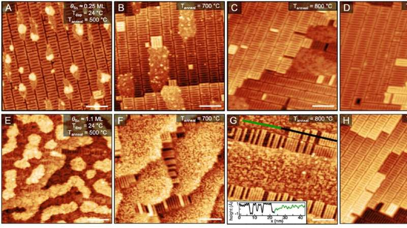
images of Sn adsorption, diffusion, incorporation, and desorption onto Nb.  Credit: Cornell University

New research from a team of scientists at the Cornell University Center for Bright Beams has made significant strides in developing new techniques to guide the growth of materials used in next-generation particle accelerators.

The study, published in the Journal of Physical Chemistry C, reveals the potential for greater control over the growth of superconducting Nb3Sn films, which could significantly reduce the cost and size of cryogenic infrastructure required for superconducting technology.

Superconducting accelerator facilities, such as those used for X-ray free-electron laser radiation, rely on niobium superconducting radio frequency (SRF) cavities to generate high-energy beams. However, the associated cryogenic infrastructure, energy consumption, and operating costs of niobium SRF cavities limit access to this technology.

To address this issue, researchers have been working to identify superconducting materials that can operate at temperatures higher than 2 Kelvin with comparable quality factors to niobium (Nb) SRF cavities. One of the most promising materials is triniobium tin (Nb3Sn), an alloy with an operating temperature of 18 Kelvin, thus reducing the need for expensive cryogenic infrastructure.

Despite theoretical and experimental advancements in the performance of Nb3Sn-coated cavities, there is still a need for a thorough understanding of how to grow higher quality Nb3Sn alloy films.

“Nb3Sn cavities are going to be the accelerators of the future,” says Ritchie Patterson, the Helen T. Edwards Professor of Physics in the College of Arts and Sciences and director of the Center for Bright Beams. “Advancing this science is only made possible through diverse collaborations—an important focus at the heart of CBB. The expertise and close collaborations between all of our partner institutions are driving this research into the future.”

Researchers capture first atomic-scale images depicting early stages of particle accelerator film formation
The metal deposition chamber. Credit: Cornell University

This new CBB research, conducted by experimental materials chemists at the University of Chicago coupled with theoretical physicists at the University of Florida, delivers the first atomic-scale images of Sn on oxidized niobium, depicting the early stages of Nb3Sn formation. This visualization of Sn adsorption and diffusion on oxidized niobium is an essential advancement in creating a mechanistic formula for optimizing the fabrication of next generation accelerator cavities.

“The quality and accelerating performance of Nb3Sn depends on many convoluted variables at play during the growth procedure,” says Sarah Willson, CBB graduate student at the University of Chicago and co-lead author of the paper along with postdoctoral scholar Rachael Farber. “We are aiming to look at the initial steps of a complicated growth process and isolate certain variables in a controlled setting.” Their atomic-level growth experiments are supported by quantum theory from graduate student Ajinkya Hire.

As Nb3Sn accelerator cavities are prepared, scientists aim to reduce impurities and contaminants from the niobium cavity to achieve a cleaner and more uniform surface. The cavity is then heated to high temperatures in the presence of an Sn vapor. This causes the Sn to diffuse into the Nb layer, forming Nb3Sn. As careful measures are taken to grow a pristine Nb3Sn film, looking closely across the cavity reveals a highly disordered, rough, polycrystalline surface—not the consistent single-crystal surface ideal for a highly controlled experiment.

Willson explains that in order to conduct this experiment, they recreate, in a way, the real-world process of cavity-making, but further surpass the temperature demands needed—heating the materials to 1630 degrees Celsius, and creating an atomically-flat niobium oxide surface to showcase the interactions of Sn, Nb, and O at the atomic level.

Observations of metal oxides are routinely performed using scanning tunneling microscopy, STM, revealing information at the atomic scale. However, the specific setup for studying Nb3Sn growth with STM is not readily available. So, Willson and Farber created one.

They designed and built a custom metal deposition chamber to deposit the Sn on the niobium surface. This technique recreates the real-world environment in which accelerator cavities are developed—with the ability to prevent surface contamination—while allowing researchers to study the deposition using STM.

Researchers capture first atomic-scale images depicting early stages of particle accelerator film formation
Credit: The Journal of Physical Chemistry C (2023). DOI: 10.1021/acs.jpcc.2c08458

“We have taken a state-of-the-art STM setup, which was not really built to study high temperature metallic growth and alloy formation, but through the funds from CBB, have added the intermetallic growth chamber that allows us to do these experiments in-situ,” says Willson, stating that using the intermetallic growth section reveals the individual Sn atoms integrating with the niobium subsurface.

“We see that even in our highly-controlled environment, the Nb surface serves as a major roadblock in preventing Sn diffusion required for Nb3Sn formation,” says Willson. “Improving Nb3Sn growth is much more than just simply developing a uniform coating layer of tin on niobium.”

This study was led by corresponding author Steven Sibener, Carl William Eisendrath Distinguished Service Professor at the University of Chicago, in collaboration with CBB faculty member Richard Hennig, Alumni Professor of Materials Science and Engineering at the University of Florida.

Sibener, a physical chemist, says that the collaboration between different areas of accelerator and non-accelerator sciences is unique in his experience, helping to lay the groundwork for advancing particle accelerators and looks forward to the promising developments of Nb3Sn.

“The collaborations that CBB sparks, the ability for surface chemists, materials engineers, accelerator physicists, and theorists to interact in this way, has certainly empowered and strengthened this research,” says Willson. “Personally, I gained a deeper understanding of how to properly navigate the challenges associated with the differing jargon, priorities, and research perspectives across scientific fields. Many chemists are interested in these types of interfacial metallic growth challenges that are encountered by engineers and physicists. This collaboration facilitated extensive interdisciplinary communication that has made conducting a study like this more comfortable and efficient.”

More information: Sarah A. Willson et al, Submonolayer and Monolayer Sn Adsorption and Diffusion Behavior on Oxidized Nb(100), The Journal of Physical Chemistry C (2023). DOI: 10.1021/acs.jpcc.2c08458

Journal information: Journal of Physical Chemistry C 

Provided by Cornell University 

New research sheds light on how harmful algal species use nitric oxide to grow

Assimilating nitric oxide
University of Delaware Associate Professor Kathyrn Coyne and Emily Healey, who received her masters at UD in marine biosciences and is currently a doctoral student at the University of Maryland School of Public Health, looked at how Heterosigma akashiwo, a globally distributed toxic species of alga, is accessing nitric oxide in the environment and how that may give it an advantage over other harmful algae species. Credit: Edward Whereat, Tammy Beeson/University of Delaware

Ever since it was discovered to be a driving force behind economically and environmentally destructive harmful algal blooms (HABs) throughout the world, researchers have been trying to discover more information about the effects of different types of nitrogen, such as nitrate or ammonium, on the proliferation of HABs.

At the University of Delaware, Kathyrn Coyne, associate professor in the School of Marine Science and Policy (SMSP), has spent several years studying HABs—including Heterosigma akashiwo, a globally distributed toxic species of alga. About 10 years ago, with funds from Delaware Sea Grant (DESG), Coyne’s lab discovered that Heterosigma akashiwo is able to use nitric oxide as a nitrogen source.

It does this by using a unique modification of an enzyme called nitrate reductase. This enzyme typically catalyzes the first step in the process of converting nitrate to ammonium, a more useable form of nitrogen. The modification of nitrate reductase in Heterosigma akashiwo allows it to use nitric oxide instead of nitrate as a source of nitrogen.

“This kind of raised a lot of questions,” Coyne said. “Where would they be accessing nitric oxide in the environment? What kind of advantage does it give Heterosigma akashiwo to have this modification in their enzyme? And how do other types of nitrogen affect its ability to use nitric oxide?”

The last question was answered in a recent article about the results of the research in Scientific Reports.

The lead author on this paper, Emily Healey, received her master’s degree at UD in marine biosciences and is a doctoral student at the University of Maryland School of Public Health. Along with Coyne, other co-authors include Joanna York, professor in the School of Marine Science and Policy and Director of DESG, and Robinson Fulweiler, professor in the Department of Earth and Environment at Boston University, as well as past members of Coyne’s lab, Stacie Flood and Patience Bock.

REU participant

Healey said that while she joined Coyne’s lab as a master’s student in the summer of 2019, she came to UD and worked with Coyne earlier as a participant in the SMSP Research Experiences for Undergraduates (REU) summer program.

“I have always been interested in microbiology and to have the opportunity to spend my summer at the beach through the REU program was pretty amazing,” Healey said. “Dr. Coyne runs a great lab. She had really helpful Ph.D. students working for her, and I just loved the work. I had never worked with algae before—I had been doing more with bacteria so it was interesting to get immersed in a different microbe.”

After her REU experience, Healey was able to come back the next year to start her master’s program. During that time, she began the research which would eventually lead to this recent publication.

Nitrate reductase

Healey said one of the goals of her research was to see if other nitrogen sources affect the activity of nitrate reductase in Heterosigma akashiwo and its ability to take up nitric oxide. For example, if there is a lot of ammonium present in an environment from agricultural sources, most species will down-regulate the nitrate reductase enzyme, which might affect Heterosigma’s ability to use nitric oxide.

“We thought if there’s a lot of ammonium, maybe Heterosigma akashiwo isn’t able to use nitric oxide as a source of nitrogen,” said Healey. “Maybe if there’s ammonium present, they simply turn off that enzyme.”

By shutting off the enzyme in the presence of ammonium, the possibility existed that Heterosigma akashiwo would be inhibited or even prevented from accessing nitric oxide and turning it into biomass.

What they found, however, was that even when there was ammonium present, providing nitric oxide to cultures of Heterosigma akashiwo actually increased activity of the enzyme so that it was able to successfully take up nitric oxide and convert it to biomass in the presence of ammonium.

To determine the effects of different types of nitrogen on nitrate reductase, the researchers conducted three experiments. They grew Heterosigma akashiwo in the lab first with only nitrate, then with only ammonium, and finally with a 50/50 mix of nitrate and ammonium.

In each of the experiments, the activity of the enzyme nitrate reductase not only increased when nitric oxide was added, but results showed that Heterosigma was incorporating nitric oxide into biomass even in the presence of ammonium.

Coyne said this ability potentially gives Heterosigma akashiwo an advantage over other species, by allowing them to access a novel source of nitrogen.

“If that were the case, then Heterosigma may be able to use nitric oxide as an alternative that other species simply don’t have access to,” said Coyne.

Nitrogen in the sediments

This ability to use nitric oxide as a nitrogen source may have something to do with how Heterosigma akashiwo moves throughout the day—as the species will move up to the surface of the water during the day and return to the bottom of the water column during the night.

“At night, Heterosigma migrates to the sediments and then during the day time, they’re photosynthetic so they move up to the surface where they are exposed to sunlight,” said Coyne. “There is a lot of data showing that nitric oxide seeps up through the sediments in some coastal areas, so we think Heterosigma may be accessing that nitric oxide as a nitrogen source at night.”

Healey said this ability to use nitric oxide may have a few implications. One would be that it gives Heterosigma akashiwo an advantage over other algae when other types of nitrogen have been depleted.

In addition, Healey said that nitric oxide should be considered a factor in bloom formation for Heterosigma akashiwo.

“People often study the effects of nitrogen on blooms and how to prevent blooms in the first place by reducing nitrate or ammonium,” said Healey. “Now, we’re saying, ‘Ok, add nitric oxide to that list because it could be important.'”

More information: Emily M. Healey et al, Effects of nitrate and ammonium on assimilation of nitric oxide by Heterosigma akashiwo, Scientific Reports (2023). DOI: 10.1038/s41598-023-27692-3

Journal information: Scientific Reports 

Provided by University of Delaware 

New frequency comb breathalyzer detects COVID-19 with excellent accuracy

JILA's Frequency Comb Breathalyzer Detects COVID-19 with Excellent Accuracy
CE-DFCS breathalyzer. (a) 
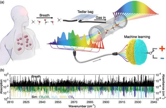
Schematic representation of the working principle of the device. An exhaled human breath sample was collected in a Tedlar bag and then loaded into an analysis chamber. The chamber was surrounded by a pair of high-reflectivity optical mirrors. A mid-infrared frequency comb laser interacted with the loaded sample and generated a broadband molecular absorption spectrum. The spectroscopy data was then used for supervised machine learning analysis to predict the binary response class for the research subject (either positive or negative). (b) Sample absorption spectrum collected from a research subject’s exhaled breath (black). Inverted in sign and plotted with different colors are four fitted species (CH3OH, H2O, HDO, and CH4) that give the most dominant absorption features. Credit: Journal of Breath Research (2023). DOI: 10.1088/1752-7163/acc6e4

JILA researchers have upgraded a breathalyzer based on Nobel Prize-winning frequency-comb technology and combined it with machine learning to detect SARS-CoV-2 infection with excellent accuracy in 170 volunteer subjects. Their achievement represents the first real-world test of the technology’s capability to diagnose disease in exhaled human breath.

Their study on this topic was published in the Journal of Breath Research.

Frequency comb technology has the potential to non-invasively diagnose more health conditions than other breath analysis techniques, while also being faster and potentially more accurate than some other medical tests. Frequency combs act as rulers for precisely measuring different colors of light, including the infrared light absorbed by biomolecules in a person’s breath.

Human breath contains more than 1,000 different trace molecules, many of which are correlated with specific health conditions. JILA’s frequency comb breathalyzer identifies chemical signatures of molecules based on exact colors and amounts of infrared light absorbed by a sample of exhaled breath.

Back in 2008, Jun Ye and colleagues at JILA demonstrated the world’s first frequency comb breathalyzer, which measured the absorption of light in the near-infrared part of the optical spectrum. In 2021 they achieved a thousandfold improvement in detection sensitivity by extending the technique to the mid-infrared spectral region, where molecules absorb light much more strongly. This enables some breath molecules to be identified at the parts-per-trillion level where those with the lowest concentrations tend to be present.

The added benefit to this study was the use of machine learning. Machine learning—a form of artificial intelligence (AI)—processes and analyzes a massive, complex mélange of data from all the breath samples as measured by 14,836 comb “teeth,” each representing a different color or frequency to create a predictive model to diagnose disease.

“Molecules increase or decrease in their concentrations when associated with specific health conditions. Machine learning analyzes this information, identifies patterns and develops reliable criteria we can use to predict a diagnosis,” said Qizhong Liang, a graduate student in the Jun Ye group, who is lead author of a new paper presenting the findings.

JILA is jointly operated by the National Institute of Standards and Technology (NIST) and the University of Colorado Boulder (CU Boulder). The research was conducted on breath samples collected from 170 CU Boulder students and staff from May 2021 to January 2022. Approximately half of the volunteers tested positive for COVID-19 with standard PCR tests. The other half of the subjects tested negative. The young study group had a median age of 23 years old, and all were above 18 years old. The general campus population was more than 90% vaccinated.

“I do think that this comb technique is superior to anything out there,” NIST/JILA Fellow Jun Ye said. “The basic point is not just the detection sensitivity, but the fact that we can generate a far greater amount of data, or breath markers, really establishing a whole new field of ‘comb breathomics’ with the help of AI. With a database, we can then use it to search and study many other physiological conditions for human beings and to help advance the future of healthcare.”

The JILA comb breathalyzer method demonstrated excellent accuracy for detecting COVID by using machine learning algorithms on absorption patterns to predict SARS-CoV-2 infection. H2O (water), HDO (semi-heavy water), H2CO (formaldehyde), NH3 (ammonia), CH3OH (methanol), and NO2 (nitrogen dioxide) were identified as discriminating molecules for detection of SARS-CoV-2 infection.

The team measured the accuracy of their results by creating a data graph comparing their predictions of COVID-19 against the PCR test results (which, it should be noted, have high but not perfect accuracy). On the graph, they computed a quantity known as the “area under the curve” (AUC). An AUC of 1, for example, would be expected for perfectly discriminating between ambient air and exhaled breath. An AUC of 0.5 would be expected for making random guesses on whether the individuals were born on odd or even months. The researchers measured an AUC of 0.849 for their COVID-19 predictions. An AUC of 0.8 or greater for medical diagnostic data is considered “excellent” accuracy.

In the future, the researchers could further increase the accuracy by expanding the spectral coverage, analyzing the patterns with more powerful AI techniques, and measuring and analyzing additional molecules, which could include the SARS-CoV-2 virus itself. Researchers would need to build a database of the specific IR colors absorbed by the virus (its spectral “fingerprint”) to potentially measure viral concentrations in the breath.

The researchers also identified significant differences in breath samples based on tobacco use and a variety of gastrointestinal symptoms such as lactose intolerance. This suggests broader capability of the technique for diagnosing different sets of diseases.

The researchers plan further studies to try to diagnose other conditions such as chronic obstructive pulmonary disease, the third-leading cause of death worldwide according to the World Health Organization. The researchers have also recently boosted the comb breathalyzer’s diagnostic power by expanding the spectral coverage to detect additional molecules. They plan to employ additional AI approaches such as deep learning to improve its disease-detection abilities. Efforts are already under way to miniaturize and simplify the technology to make it portable and easy to use in hospitals and other care settings.

Ye said there is interest from the medical community in seeing the comb breathalyzer developed further and commercialized. Approval by the U.S. Food and Drug Administration (FDA) would be needed before the technology could be used in medical settings.

The most prevalent analytical technique in breath research now is gas chromatography combined with mass spectrometry, which can detect hundreds of exhaled molecules but works slowly, typically requiring tens of minutes. Its use of chemical process also unavoidably alters breath components and presents analytical challenges to identify breath profiles accurately. Frequency comb technology measures breath molecules in a non-destructive and real time manner and can promote a more accurate and repeatable determination of exhaled breath contents.

More information: Qizhong Liang et al, Breath analysis by ultra-sensitive broadband laser spectroscopy detects SARS-CoV-2 infection, Journal of Breath Research (2023). DOI: 10.1088/1752-7163/acc6e4

Journal information: Journal of Breath Research 

Provided by National Institute of Standards and Technology 

This story is republished courtesy of NIST. Read the original story here.

New atomic-scale understanding of catalysis could unlock massive energy savings

atomic
Credit: Pixabay/CC0 Public Domain

In an advance they consider a breakthrough in computational chemistry research, University of Wisconsin–Madison chemical engineers have developed model of how catalytic reactions work at the atomic scale. This understanding could allow engineers and chemists to develop more efficient catalysts and tune industrial processes—potentially with enormous energy savings, given that 90% of the products we encounter in our lives are produced, at least partially, via catalysis.

Catalyst materials accelerate chemical reactions without undergoing changes themselves. They are critical for refining petroleum products and for manufacturing pharmaceuticals, plastics, food additives, fertilizers, green fuels, industrial chemicals and much more.

Scientists and engineers have spent decades fine-tuning catalytic reactions—yet because it’s currently impossible to directly observe those reactions at the extreme temperatures and pressures often involved in industrial-scale catalysis, they haven’t known exactly what is taking place on the nano and atomic scales. This new research helps unravel that mystery with potentially major ramifications for industry.

In fact, just three catalytic reactions—steam-methane reforming to produce hydrogen, ammonia synthesis to produce fertilizer, and methanol synthesis—use close to 10% of the world’s energy.

“If you decrease the temperatures at which you have to run these reactions by only a few degrees, there will be an enormous decrease in the energy demand that we face as humanity today,” says Manos Mavrikakis, a professor of chemical and biological engineering at UW–Madison who led the research. “By decreasing the energy needs to run all these processes, you are also decreasing their environmental footprint.”

Mavrikakis and postdoctoral researchers Lang Xu and Konstantinos G. Papanikolaou along with graduate student Lisa Je published news of their advance in the April 7, 2023 issue of the journal Science.

In their research, the UW–Madison engineers develop and use powerful modeling techniques to simulate catalytic reactions at the atomic scale. For this study, they looked at reactions involving transition metal catalysts in nanoparticle form, which include elements like platinum, palladium, rhodium, copper, nickel, and others important in industry and green energy.

According to the current rigid-surface model of catalysis, the tightly packed atoms of transition metal catalysts provide a 2D surface that chemical reactants adhere to and participate in reactions. When enough pressure and heat or electricity is applied, the bonds between atoms in the chemical reactants break, allowing the fragments to recombine into new chemical products.

“The prevailing assumption is that these metal atoms are strongly bonded to each other and simply provide ‘landing spots’ for reactants. What everybody has assumed is that metal-metal bonds remain intact during the reactions they catalyze,” says Mavrikakis. “So here, for the first time, we asked the question, ‘Could the energy to break bonds in reactants be of similar amounts to the energy needed to disrupt bonds within the catalyst?'”

According to Mavrikakis’s modeling, the answer is yes. The energy provided for many catalytic processes to take place is enough to break bonds and allow single metal atoms (known as adatoms) to pop loose and start traveling on the surface of the catalyst. These adatoms combine into clusters, which serve as sites on the catalyst where chemical reactions can take place much easier than the original rigid surface of the catalyst.

Using a set of special calculations, the team looked at industrially important interactions of eight transition metal catalysts and 18 reactants, identifying energy levels and temperatures likely to form such small metal clusters, as well as the number of atoms in each cluster, which can also dramatically affect reaction rates.

Their experimental collaborators at the University of California, Berkeley, used atomically-resolved scanning tunneling microscopy to look at carbon monoxide adsorption on nickel (111), a stable, crystalline form of nickel useful in catalysis. Their experiments confirmed models that showed various defects in the structure of the catalyst can also influence how single metal atoms pop loose, as well as how reaction sites form.

Mavrikakis says the new framework is challenging the foundation of how researchers understand catalysis and how it takes place. It may apply to other non-metal catalysts as well, which he will investigate in future work. It is also relevant to understanding other important phenomena, including corrosion and tribology, or the interaction of surfaces in motion.

“We’re revisiting some very well-established assumptions in understanding how catalysts work and, more generally, how molecules interact with solids,” Mavrikakis says.

Manos Mavrikakis is Ernest Micek Distinguished Chair, James A. Dumesic Professor, and Vilas Distinguished Achievement Professor in Chemical and Biological Engineering at the University of Wisconsin–Madison. Other authors include Barbara A.J. Lechner of the Technical University of Munich, and Gabor A. Somorjai and Miquel Salmeron of Lawrence Berkeley National Laboratory and the University of California, Berkeley.

More information: Lang Xu et al, Formation of active sites on transition metals through reaction-driven migration of surface atoms, Science (2023). DOI: 10.1126/science.add0089www.science.org/doi/10.1126/science.add0089

Journal information: Science 

Provided by University of Wisconsin-Madison 

Scientists use peroxide to peer into metal oxide reactions

Scientists use peroxide to peer into metal oxide reactions
Lab Based Ambient Pressure X-ray Photoelectron Spectroscopy (XPS) Instrument at CFN. Credit: Brookhaven National Laboratory

Researchers at Binghamton University led research partnering with the Center for Functional Nanomaterials (CFN)—a U.S. Department of Energy (DOE) Office of Science User Facility at Brookhaven National Laboratory—to get a better look at how peroxides on the surface of copper oxide promote the oxidation of hydrogen but inhibit the oxidation of carbon monoxide, allowing them to steer oxidation reactions. They were able to observe these quick changes with two complimentary spectroscopy methods that have not been used in this way. The results of this work have been published in the journal Proceedings of the National Academy of Sciences (PNAS).

“Copper is one of the most studied and relevant surfaces, both in catalysis and in corrosion science,” explained Anibal Boscoboinik, materials scientist at CFN. “So many mechanical parts that are used in industry are made of copper, so trying to understand this element of the corrosion processes is very important.”

“I’ve always liked looking at copper systems,” said Ashley Head also a materials scientist at CFN. “They have such interesting properties and reactions, some of which are really striking.”

Gaining a better understanding of oxide catalysts gives researchers more control of the chemical reactions they produce, including solutions for clean energy. Copper, for example, can catalytically form and convert methanol into valuable fuels, so being able to control the amount of oxygen and number of electrons on copper is a key step to efficient chemical reactions.

Peroxide as a proxy

Peroxides are chemical compounds that contain two oxygen atoms linked by shared electrons. The bond in peroxides is fairly weak, allowing other chemicals to alter its structure, which makes them very reactive. In this experiment, scientists were able to alter the redox steps of catalytic oxidation reactions on an oxidized copper surface (CuO) by identifying the makeup of peroxide species formed with different gases: O2 (oxygen), H2 (hydrogen), and CO (carbon monoxide).

Redox is a combination of reduction and oxidation. In this process, the oxidizing agent gains an electron and the reducing agent loses an electron. When comparing these different peroxide species and how these steps played out, researchers found that a surface layer of peroxide significantly enhanced CuO reducibility in favor of H2 oxidation. They also found that, on the other hand, it acted as an inhibitor to suppress CuO reduction against CO (carbon monoxide) oxidation. They found that this opposite effect of the peroxide on the two oxidation reactions stems from the modification of the surface sites where the reaction takes place.

By finding these bonding sites and learning how they promote or inhibit oxidation, scientists can use these gases to gain more control of how these reactions play out. In order to tune these reactions though, scientists had to get a clear look at what was happening.

Scientists use peroxide to peer into metal oxide reactions
Binding energy and location of peroxide (OO) formation on Copper Oxide (CuO). Credit: Proceedings of the National Academy of Sciences (PNAS)

The right tools for the job

Studying this reaction in situ was important to the team, since peroxides are very reactive and these changes happen fast. Without the right tools or environment, it’s hard to catch such a limited moment on the surface.

Peroxide species on copper surfaces were never observed using in-situ infrared (IR) spectroscopy in the past. With this technique, researchers use infrared radiation to get a better understanding of a material’s chemical properties by looking at the way the radiation is absorbed or reflected under reaction conditions. In this experiment, scientists were able to differentiate “species” of peroxide, with very slight variations in the oxygen they were carrying, which would have otherwise been very hard to identify on a metal oxide surface.

“I got really excited when I was looking up the infrared spectra of these peroxide species on a surface and seeing that there weren’t many publications. It was exciting that we could see these differences using a technique that’s not widely applied to these kind of species,” recalled Head.

IR spectroscopy on its own wasn’t enough to be sure though, which is why the team also used another spectroscopy technique called ambient pressure X-ray Photoelectron Spectroscopy (XPS). XPS uses lower energy X-rays to kick electrons out of the sample. The energy of these electrons gives scientists clues about the chemical properties of atoms in the sample. Having both techniques available through the CFN User Program was key to making this research possible.

“One of the things that we pride ourselves in is the instruments that we have and modified here,” said Boscoboinik. “Our instruments are connected, so users can move the sample in a controlled environment between these two techniques and study them in situ to get complementary information. In most other circumstances, a user would have to take the sample out to go to a different instrument, and that change of environment could alter its surface.”

“A nice feature of CFN lies not only in its state-of-the-art facilities for science, but also the opportunities it provides to train young researchers,” said Guangwen Zhou professor at the Thomas J. Watson College of Engineering and Applied Science’s Department of Mechanical Engineering and the Materials Science program at Binghamton University. “Each of the students involved have benefited from extensive, hands-on experience in the microscopy and spectroscopy tools available at CFN.”

This work was accomplished with the contributions of four Ph.D. students in Zhou’s group: Yaguang Zhu and Jianyu Wang, the first co-authors of this paper, and Shyam Patel and Chaoran Li. All of these students are early in their career, having just earned their PhDs in 2022.

Future findings

The results of this study may apply to other types of reactions and other catalysts besides copper. These findings and the processes and techniques that led scientists there could find their ways into related research. Metal oxides are widely used as catalysts themselves or components in catalysts. Tuning peroxide formation on other oxides could be a way to block or enhance surface reactions during other catalytic processes.

“I’m involved in some other projects related to copper and copper oxides, including transforming carbon dioxide to methanol to use as a fuel for clean energy,” said Head. “Looking at these peroxides on the same surface that I use has the potential to make an impact on other projects using copper and other metal oxides.”

More information: Yaguang Zhu et al, Tuning the surface reactivity of oxides by peroxide species, Proceedings of the National Academy of Sciences (2023). DOI: 10.1073/pnas.2215189120

Journal information: Proceedings of the National Academy of Sciences 

Provided by Brookhaven National Laboratory