Scientists design superdiamonds with theoretically predicted hexagonal crystal structure

The brilliantly shiny diamond is more than just pretty; it’s one of the hardest minerals on Earth, with a name derived from the Greek word adámas, meaning unbreakable. Scientists have now engineered a harder form of diamond known as bulk hexagonal diamond (HD)—a crystalline structure that has been theorized for over half a century to have physical properties superior to those of conventional diamond.

In a study published in Nature, researchers from China synthesized bulk hexagonal diamond, ranging from 100-µm-sized to mm-sized, with a highly ordered structure by compressing and heating high-quality graphite single crystals under pressure conditions as uniform as possible.

The designed material, which was recoverable under ambient conditions, unveiled the previously elusive structural world of HD, opening new avenues for exploring its potential as a technologically superior material.

Apart from being a dazzling element in jewelry, diamonds, due to their unmatched chemical and physical properties, are sought after in a wide range of applications, including biosensors, quantum computing, and industrial processes as superabrasives and drilling bits.

These properties arise from its unique cubic atomic structure, where each carbon atom is bonded to four other carbon atoms via strong sp3 bonds, giving rise to a rigid tetrahedral network.

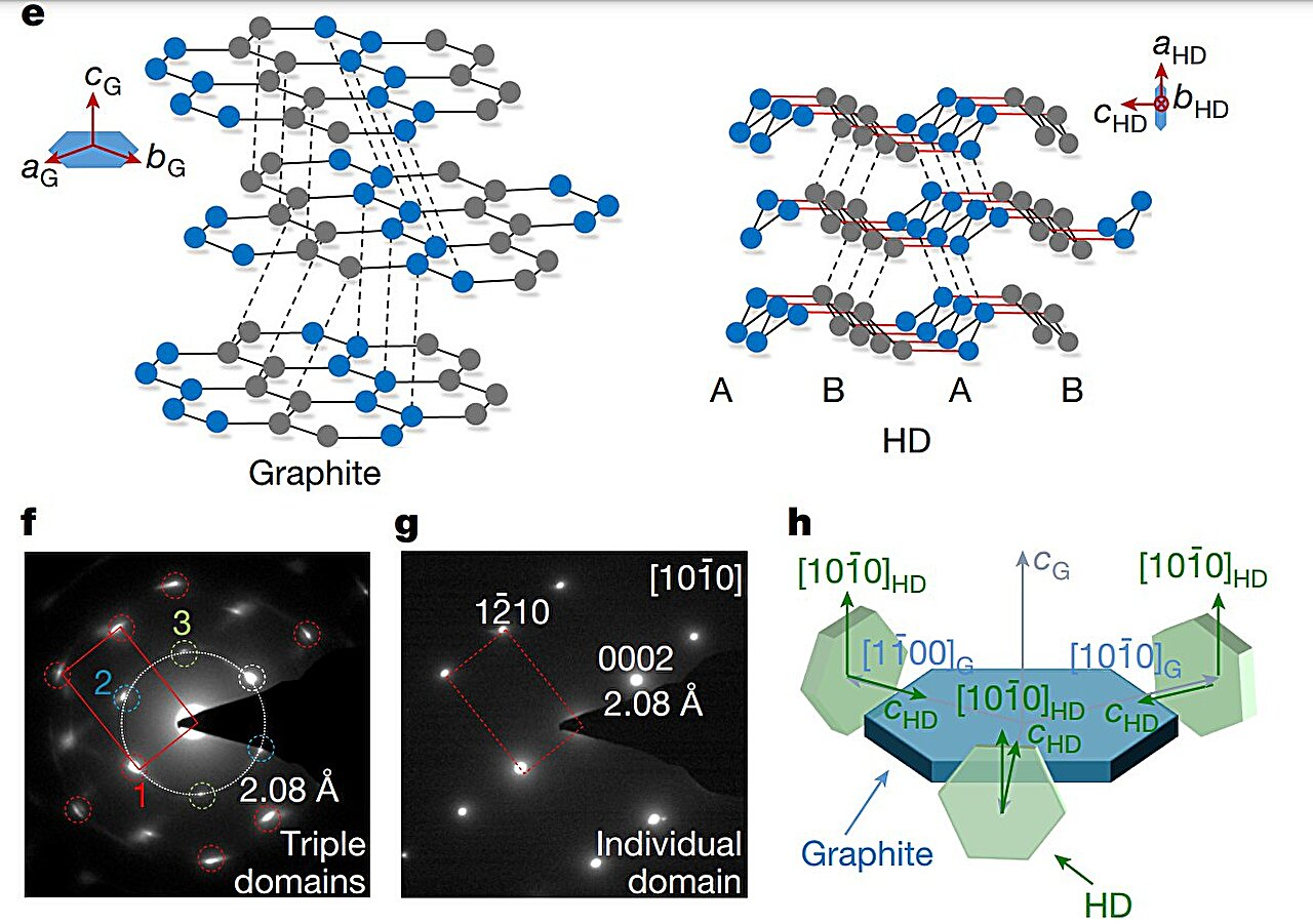
These structures then form honeycomb layers that stack on top of each other to build cubic diamond crystals. The natural cleavage planes created by these layered structures is also the reason behind diamond’s strength limit, something that research suggests can be rectified by selectively strengthening and shortening the interlayer bonds. This change, however, would transform the cubic diamond into a hexagonal crystal symmetry.HRTEM images and corresponding SAD patterns of the bulk HD sample were acquired along different zone axes. Credit: Nature (2025). DOI: 10.1038/s41586-025-09343-x

Similar transformations aren’t unseen in nature, for instance, HD, also known as lonsdaleite, is found in Canyon Diablo meteorite, where it was named.

2022 study suggested that the high temperatures and shock compressions created during the meteor impact turn graphite in the meteorite into HD.

Scientists have attempted to emulate similar explosive and high-pressure conditions in laboratories to produce artificial lonsdaleite but most samples are too small and often impure, making it difficult to isolate and study the true properties of HD.

Now, the researchers have successfully synthesized a highly ordered and nearly pure bulk HD, which was recovered intact under ambient conditions. Starting from single-crystal hexagonal graphite, they created a controlled quasi-hydrostatic environment—applying pressure as uniformly as possible to minimize stress variations—using both a diamond anvil cell (DAC) and a large-volume multi-anvil press.

The bulk sample consisted of a threefold intergrowth of densely packed crystals, each approximately 100 nanometers in size, primarily composed of HD with minor imperfections, including traces of cubic diamond.Pure sp3 bonding state and the excellent mechanical properties of bulk HD. Credit: Nature (2025). DOI: 10.1038/s41586-025-09343-x

Investigating the molecular-level mechanisms via spectroscopy revealed a complete conversion of sp2 bonds into sp3 bonds, indicating a direct crystallographic transformation from graphite to diamond.

They also found that the interlayer bonds of HD were stronger and shorter than those in conventional cubic diamond, which was reflected in the Vickers hardness test—which measures a material’s resistance to plastic deformation—where the values for HD were slightly higher.

The researchers note that this study unambiguously demonstrates the existence of HD as a bona fide phase of carbon with superior hardness, similar to that of cubic diamond. They believe that experimenting with levels of graphite precursor purification and fine-tuning the pressure–temperature conditions can lead to the creation of even higher-quality HD with superior properties.

AI-guided search uncovers new molecules for stronger, longer-lasting plastics

A new strategy for strengthening polymer materials could lead to more durable plastics and cut down on plastic waste, according to researchers at MIT and Duke University.

Using machine learning, the researchers identified crosslinker molecules that can be added to polymer materials, allowing them to withstand more force before tearing. These crosslinkers belong to a class of molecules known as mechanophores, which change their shape or other properties in response to mechanical force.

“These molecules can be useful for making polymers that would be stronger in response to force. You apply some stress to them, and rather than cracking or breaking, you instead see something that has higher resilience,” says Heather Kulik, the Lammot du Pont Professor of Chemical Engineering at MIT, who is also a professor of chemistry and the senior author of the study.

The crosslinkers that the researchers identified in this study are iron-containing compounds known as ferrocenes, which until now had not been broadly explored for their potential as mechanophores. Experimentally evaluating a single mechanophore can take weeks, but the researchers showed that they could use a machine-learning model to dramatically speed up this process.

MIT postdoc Ilia Kevlishvili is the lead author of the open-access paper, which appeared in ACS Central Science.

Other authors include Jafer Vakil, a Duke graduate student; David Kastner and Xiao Huang, both MIT graduate students; and Stephen Craig, a professor of chemistry at Duke.

The weakest link
Mechanophores are molecules that respond to force in unique ways, typically by changing their color, structure, or other properties. In the new study, the MIT and Duke team wanted to investigate whether they could be used to help make polymers more resilient to damage.

The new work builds on a 2023 study by Craig and Jeremiah Johnson, the A. Thomas Guertin Professor of Chemistry at MIT, and their colleagues.

In that work, the researchers found that, surprisingly, incorporating weak crosslinkers into a polymer network can make the overall material stronger.

When materials with these weak crosslinkers are stretched to the breaking point, any cracks propagating through the material try to avoid the stronger bonds and go through the weaker bonds instead. This means the crack has to break more bonds than it would if all of the bonds were the same strength.

To find new ways to exploit that phenomenon, Craig and Kulik joined forces to try to identify mechanophores that could be used as weak crosslinkers.

“We had this new mechanistic insight and opportunity, but it came with a big challenge: of all possible compositions of matter, how do we zero in on the ones with the greatest potential?” Craig says.

“Full credit to Heather and Ilia for both identifying this challenge and devising an approach to meet it.”

Discovering and characterizing mechanophores is a difficult task that requires either time-consuming experiments or computationally intense simulations of molecular interactions. Most of the known mechanophores are organic compounds, such as cyclobutane, which was used as a crosslinker in the 2023 study.

In the new study, the researchers wanted to focus on molecules known as ferrocenes, which are believed to hold potential as mechanophores. Ferrocenes are organometallic compounds that have an iron atom sandwiched between two carbon-containing rings. Those rings can have different chemical groups added to them, which alter their chemical and mechanical properties.

Many ferrocenes are used as pharmaceuticals or catalysts, and a handful are known to be good mechanophores, but most have not been evaluated for that use. Experimental tests on a single potential mechanophore can take several weeks, and computational simulations, while faster, still take a couple of days. Evaluating thousands of candidates using these strategies is a daunting task.

Realizing that a machine-learning approach could dramatically speed up the characterization of these molecules, the MIT and Duke team decided to use a neural network to identify ferrocenes that could be promising mechanophores.

They began with information from a database known as the Cambridge Structural Database, which contains the structures of 5,000 different ferrocenes that have already been synthesized.

“We knew that we didn’t have to worry about the question of synthesizability, at least from the perspective of the mechanophore itself. This allowed us to pick a really large space to explore with a lot of chemical diversity that would also be synthetically realizable,” Kevlishvili says.

First, the researchers performed computational simulations for about 400 of these compounds, allowing them to calculate how much force is necessary to pull atoms apart within each molecule. For this application, they were looking for molecules that would break apart quickly, as these weak links could make polymer materials more resistant to tearing.

Then they used this data, along with information on the structure of each compound, to train a machine-learning model. This model was able to predict the force needed to activate the mechanophore, which in turn influences resistance to tearing, for the remaining 4,500 compounds in the database, plus an additional 7,000 compounds that are similar to those in the database but have some atoms rearranged.

The researchers discovered two main features that seemed likely to increase tear resistance. One was interactions between the chemical groups that are attached to the ferrocene rings. Additionally, the presence of large, bulky molecules attached to both rings of the ferrocene made the molecule more likely to break apart in response to applied forces.

While the first of these features was not surprising, the second trait was not something a chemist would have predicted beforehand, and could not have been detected without AI, the researchers say. “This was something truly surprising,” Kulik says.

Tougher plastics
Once the researchers identified about 100 promising candidates, Craig’s lab at Duke synthesized a polymer material incorporating one of them, known as m-TMS-Fc. Within the material, m-TMS-Fc acts as a crosslinker, connecting the polymer strands that make up polyacrylate, a type of plastic.

By applying force to each polymer until it tore, the researchers found that the weak m-TMS-Fc linker produced a strong, tear-resistant polymer. This polymer turned out to be about four times tougher than polymers made with standard ferrocene as the crosslinker.

“That really has big implications, because if we think of all the plastics that we use and all the plastic waste accumulation, if you make materials tougher, that means their lifetime will be longer. They will be usable for a longer period of time, which could reduce plastic production in the long term,” Kevlishvili says.

The researchers now hope to use their machine-learning approach to identify mechanophores with other desirable properties, such as the ability to change color or become catalytically active in response to force. Such materials could be used as stress sensors or switchable catalysts, and they could also be useful for biomedical applications such as drug delivery.

In those studies, the researchers plan to focus on ferrocenes and other metal-containing mechanophores that have already been synthesized but whose properties are not fully understood.

“Transition metal mechanophores are relatively underexplored, and they’re probably a little bit more challenging to make,” Kulik says. “This computational workflow can be broadly used to enlarge the space of mechanophores that people have studied.”

Cool new imaging method shows elemental distributions of nanomaterials in frozen solvents

Cryo-transmission electron microscopy (cryo-TEM) allows us to observe samples in a preserved state that is close to their native form, making it a highly effective way to examine biological samples. This technique provides information on the size, shape, and dispersion of samples within a frozen solvent. However, there is another crucial piece of information that has not been accurately visualized in organic samples using this technique yet: elemental composition.

Energy-filtered TEM (EF-TEM) can determine the elemental composition by using electron energy loss spectroscopy (EELS), but it has its shortcomings. The conventional EELS/EF-TEM method can significantly damage the sample and usually exhibits drift—which results in a blurry image. Furthermore, conventional observations have primarily been applied to metals or large-area samples, rather than to organic nanomaterials.

To overcome these limitations, researchers at Tohoku University developed a novel elemental mapping technique using cryo-EELS/EF-TEM, enabling simultaneous visualization of both the structure and elemental distribution of nanomaterials in frozen solvents. This new approach allows high-resolution analysis of organic and bio-related materials. The findings were published in Analytical Chemistry on July 31, 2025.

The team found that plasmon signals from ice strongly increased the background signal—an undesirable effect since they are focused on the organic sample and aren’t interested in signals from the ice itself. To remedy this, the team developed a new imaging method combining the 3-window method for accurate background correction with drift compensation techniques. Additionally, a new program was created for the “ParallEM” electron microscope control system, allowing easy control of energy shift during imaging.Elemental mapping of hydroxyapatite particles using cryo-EELS imaging. (Left) The conventional cryo-TEM image shows the size, shape, and dispersion of the hydroxyapatite particles. (Center and right) Calcium and phosphorus—constituent elements of hydroxyapatite—are successfully detected and visualized. Credit: Daisuke Unabara et al

Using this technique, the team successfully visualized silicon distribution in silica nanoparticles within a frozen solvent, alongside their structure and dispersion. The smallest particle size detectable via elemental mapping was determined to be around 10 nm.

The method was also applied to hydroxyapatite particles, a major component of bones and teeth, revealing calcium and phosphorus distributions—key biological elements.

This technique is expected to enable advanced analysis of various materials, including biomaterials, medical materials, food, catalysts, and inks. As such, it can contribute to research across fields from life sciences to materials science.

High-quality crystals enable new insights into structure–property relationships and multifunctionality

Researchers at Kumamoto University and Nagoya University have developed a new class of two-dimensional (2D) metal-organic frameworks (MOFs) using triptycene-based molecules, marking a breakthrough in the quest to understand and enhance the physical properties of these promising materials. The work is published in the Journal of the American Chemical Society.

This innovation opens new possibilities for multifunctional applications in gas/molecular sensors, electrochemical energy storage, and spintronic devices.

Two-dimensional (2D) conductive metal-organic frameworks (MOFs) have drawn increasing attention for their unique properties—like high electron and proton conductivity and unusual magnetic behaviors—that set them apart from conventional MOFs. However, the field has long been held back by challenges in growing large, high-quality crystals and a lack of clarity about how molecular structures relate to material performance.

To tackle these issues, Associate Professor Zhongyue Zhang from the Faculty of Advanced Science and Technology at Kumamoto University, in collaboration with Professor Kunio Awaga’s team at Nagoya University at the time of the research, turned to triptycene-based linkers.

Unlike traditional flat, π-conjugated ligands that promote fast crystal growth and stacking, triptycene has a rigid 3D shape that suppresses interlayer interactions. This allows crystals to grow more slowly and reach larger sizes, making them suitable for detailed structural and functional studies.

Using a slow diffusion method in sealed glass tubes—rather than standard solvothermal techniques—the team successfully synthesized two new MOFs: Cu3(TripH2)2 and Cu3(TripMe2)2.

These reached crystal sizes exceeding 0.3 mm, sufficient for single-crystal X-ray diffraction and precise measurements of electronic, magnetic, and proton transport properties.

Structural analysis revealed that the catechol groups coordinating the metal ions remain fully protonated—an unusual and experimentally verified feature that stabilizes the layered framework via hydrogen bonding.

While previous theories suggested protonation might affect electronic properties, this study provides the first experimental evidence of such protonated states in MOFs.

Measurements on these large single crystals showed high directional conductivities, with electron and proton transport significantly stronger along the vertical (a-axis) direction. The data suggest a possible cooperative mechanism for charge and proton hopping between different arms of the triptycene units.

Electron paramagnetic resonance (EPR) and magnetization studies further revealed one-dimensional antiferromagnetic coupling along the same axis, enabled by interlayer hydrogen bonds. This is in sharp contrast to the frustrated in-plane magnetic behavior seen in other 2D MOFs.

Because these materials exhibit strong electronic and magnetic correlations in the interlayer direction—despite having no continuous structural connections there—the researchers propose a new term to describe them: “2.5-dimensional” (2.5-D) MOFs.

“This study demonstrates how a simple change in molecular geometry can overcome longstanding barriers in MOF research,” said Professor Zhongyue Zhang of Kumamoto University.

“By enabling high-quality single crystals, we not only clarified fundamental structure–property relationships but also unlocked new potential for next-generation MOFs in real-world devices.”

The findings pave the way for further developments in MOF-based technologies, including zinc-ion batteries, molecular sensors, and quantum information systems.

Polymer-protected DNA sensors enable two-month storage for 50-cent disease diagnostics

Using an inexpensive electrode coated with DNA, MIT researchers have designed disposable diagnostics that could be adapted to detect a variety of diseases, including cancer or infectious diseases such as influenza and HIV.

These electrochemical sensors make use of a DNA-chopping enzyme found in the CRISPR gene-editing system. When a target such as a cancerous gene is detected by the enzyme, it begins shearing DNA from the electrode nonspecifically, like a lawnmower cutting grass, altering the electrical signal produced.

One of the main limitations of this type of sensing technology is that the DNA that coats the electrode breaks down quickly, so the sensors can’t be stored for very long and their storage conditions must be tightly controlled, limiting where they can be used. In a new study, MIT researchers stabilized the DNA with a polymer coating, allowing the sensors to be stored for up to two months, even at high temperatures. After storage, the sensors were able to detect a prostate cancer gene that is often used to diagnose the disease.

The DNA-based sensors, which cost only about 50 cents to make, could offer a cheaper way to diagnose many diseases in low-resource regions, says Ariel Furst, the Paul M. Cook Career Development Assistant Professor of Chemical Engineering at MIT and the senior author of the study.

“Our focus is on diagnostics that many people have limited access to, and our goal is to create a point-of-use sensor. People wouldn’t even need to be in a clinic to use it. You could do it at home,” Furst says.

MIT graduate student Xingcheng Zhou is the lead author of the paper published in the journal ACS Sensors. Other authors of the paper are MIT undergraduate Jessica Slaughter, Smah Riki ’24, and graduate student Chao Chi Kuo.

An inexpensive sensor
Electrochemical sensors work by measuring changes in the flow of an electric current when a target molecule interacts with an enzyme. This is the same technology that glucose meters use to detect concentrations of glucose in a blood sample.

The electrochemical sensors developed in Furst’s lab consist of DNA adhered to an inexpensive gold leaf electrode, which is laminated onto a sheet of plastic. The DNA is attached to the electrode using a sulfur-containing molecule known as a thiol.

In a 2021 study, Furst’s lab showed that they could use these sensors to detect genetic material from HIV and human papillomavirus (HPV). The sensors detect their targets using a guide RNA strand, which can be designed to bind to nearly any DNA or RNA sequence. The guide RNA is linked to an enzyme called Cas12, which cleaves DNA nonspecifically when it is turned on and is in the same family of proteins as the Cas9 enzyme used for CRISPR genome editing.

If the target is present, it binds to the guide RNA and activates Cas12, which then cuts the DNA adhered to the electrode. That alters the current produced by the electrode, which can be measured using a potentiostat (the same technology used in handheld glucose meters).

“If Cas12 is on, it’s like a lawnmower that cuts off all the DNA on your electrode, and that turns off your signal,” Furst says.

In previous versions of the device, the DNA had to be added to the electrode just before it was used, because DNA doesn’t remain stable for very long. In the new study, the researchers found that they could increase the stability of the DNA by coating it with a polymer called polyvinyl alcohol (PVA).

This polymer, which costs less than 1 cent per coating, acts like a tarp that protects the DNA below it. Once deposited onto the electrode, the polymer dries to form a protective thin film.

“Once it’s dried, it seems to make a very strong barrier against the main things that can harm DNA, such as reactive oxygen species that can either damage the DNA itself or break the thiol bond with the gold and strip your DNA off the electrode,” Furst says.

New fluorescent probe enables rapid, visible detection of harmful pesticide residues

A team of researchers led by Prof. Jiang Changlong from the Hefei Institutes of Physical Science of the Chinese Academy of Sciences has developed a fast and simple way to detect harmful pesticide residues, with results visible to the naked eye in just 10 seconds. The study was published in Analytical Chemistry.

While effective against pests, overexposure to pyrethroids can cause health issues such as dizziness and breathing problems. Detecting them usually requires lab equipment and time-consuming procedures. But this new probe offers a much faster and easier solution.

“We created a new fluorescent probe that lights up in different colors when it comes into contact with pyrethroids,” said Liu Anqi, a member of the team. “It’s a widely used type of insecticide found in many household and agricultural products.”

The probe consists of a special fluorescent dye combined with a common protein to form a molecular complex. Under ultraviolet light, the complex emits a yellow glow. When it encounters pyrethroid molecules, the fluorescence shifts to green—an easily distinguishable change visible to the unaided eye within 10 seconds.

Tests showed that the probe is capable of detecting a broad range of pyrethroid concentrations with high sensitivity. For practical use, the researchers also developed a portable system that users can photograph the glowing sample with a smartphone and estimate pesticide levels based on image color analysis, enabling rapid field testing in kitchens, markets, or agricultural settings.

To detect pesticide vapors—such as those emitted by mosquito coils—the researchers went a step further. They embedded the fluorescent probe into a lightweight, sponge-like aerogel material. This aerogel traps airborne pesticides and shows a visible color change from orange to green. It’s one of the first tools of its kind designed for gas-phase pesticide detection.

This breakthrough opens new doors for fast, accessible pesticide monitoring, helping improve food safety and reduce health risks.

AI-designed inhibitor targets key enzyme to fight prostate cancer drug resistance

Prostate cancer is one of the most common malignancies in men globally. Hormonal therapies targeting the androgen–androgen receptor axis have significantly delayed disease progression. However, drug resistance remains inevitable, and new therapeutic targets and strategies are required to overcome androgen receptor pathway inhibitor (ARPI) resistance.

In a study published in the Proceedings of the National Academy of Sciences, Dr. Li Zhenfei’s team from the Center for Excellence in Molecular Cell Science (Shanghai Institute of Biochemistry and Cell Biology) of the Chinese Academy of Sciences (CAS), Dr. Hu Youhong’s team and Liu Jia’s team from the Shanghai Institute of Materia Medica of CAS, and Dr. Ren Ruobing’s team from Fudan University designed a novel 3βHSD1 inhibitor, HEAL-116, which has superior enzymatic activity and favorable pharmacokinetic properties, providing a new strategy for prostate cancer treatment and overcoming resistance to second-generation ARPIs.

Previous research by Dr. Li’s team has identified the metabolic enzyme 3βHSD1 as a key driver for prostate cancer progression, which regulates the metabolism of androgen, progesterone, and abiraterone, mediating resistance to ARPIs, and has also identified biochanin A (BCA) as a potent 3βHSD1 inhibitor to suppress the development of prostate cancer in cell lines, mouse models, and even patients. However, BCA’s low oral bioavailability hindered its clinical translation.

In this study, the researchers constructed a high-precision structure model of 3βHSD1 by integrating AlphaFold2 protein structure predictions, molecular dynamics simulations, quantum chemistry calculations and other techniques, and revealed its unique catalytic mechanism and substrate-binding pocket characteristics.

Through systematic optimization of BCA’s molecular geometry and charge distribution, the researchers developed HEAL-116, a highly specific inhibitor with enhanced binding affinity and improved oral bioavailability via hydrophilic group modifications.

In vivo and in vitro experiments showed that HEAL-116 potently suppressed 3βHSD1 activity and inhibited the growth of prostate cancer xenografts, when used alone or in combination with ARPIs. The specificity of HEAL-116 was also evaluated, showing no significant effects on transcriptome and kinome.

This study validates the artificial intelligence-driven rational drug design strategy. It provides a new strategy to overcome prostate cancer drug resistance, and promotes the clinical application of 3βHSD1-targeted therapy.

Novel cross-linker streamlines protein complex analysis in living cells

Proteins have specific biological functions in cells through conformational changes and interactions. Therefore, precise, in situ analysis of protein complex changes is essential for understanding cellular functions, uncovering disease mechanisms, and identifying potential drug targets.

In vivo cross-linking mass spectrometry (XL-MS) has emerged as a powerful technique to study protein complexes in living cells. However, during the enrichment of cross-linked peptides, complicated steps cause considerable sample loss, which limits the analysis of limited samples and hampers the reproducibility in quantitative analysis.

In a study published in Angewandte Chemie International Edition, a team led by Prof. Zhang Lihua from the Dalian Institute of Chemical Physics (DICP) of the Chinese Academy of Sciences developed a novel dimethylpiperidine-based cross-linker that enables one-step enrichment and quantitative analysis of protein complexes.

Using dimethylpiperidine as the enrichment handle, the researchers designed a cross-linker, 2,6-dimethylpiperidine disuccinimidyl tridecanoate (DPST). DPST exhibited cell membrane permeability, enrichment capability, and quantitative functionality.

By leveraging tandem mass tag antibodies, DPST enabled one-step enrichment and reversible elution of cross-linked peptides, eliminating sample loss from traditional multi-step processes. This allowed in vivo XL-MS analysis using as few as 10,000 cells.

Moreover, DPST supported light and heavy isotope labeling at the cellular level, and improved signal-to-noise ratio via MS2 quantitative reporter ions without increasing spectral complexity.

Using this novel cross-linker, researchers successfully mapped the protein interaction network in primary neurons derived from a single-embryo mouse. They also achieved quantitative detection of transient and weak interactions within dynamic liquid-liquid phase separation environments.

“By addressing key limitations in in vivo cross-linking proteomics, DPST provides a powerful solution for both qualitative and quantitative XL-MS analysis, with strong potential to drive advances in biomedical research and drug discovery,” said Prof. Zhang.

A new platform for developing advanced metals at scale

Companies building next-generation products and breakthrough technologies are often limited by the physical constraints of traditional materials. In aerospace, defense, energy, and industrial tooling, pushing those constraints introduces possible failure points into the system, but companies don’t have better options, given that producing new materials at scale involves multiyear timelines and huge expenses.

Foundation Alloy wants to break the mold. The company, founded by a team from MIT, is capable of producing a new class of ultra-high-performance metal alloys using a novel production process that doesn’t rely on melting raw materials. The company’s solid-state metallurgy technology, which simplifies the development and manufacturing of next-generation alloys, was developed over many years of research by former MIT professor Chris Schuh and collaborators.

“This is an entirely new approach to making metals,” says CEO Jake Guglin MBA ’19, who co-founded Foundation Alloy with Schuh, Jasper Lienhard ’15, Ph.D. ’22, and Tim Rupert Ph.D. ’11. “It gives us a broad set of rules on the materials engineering side that allows us to design a lot of different compositions with previously unattainable properties. We use that to make products that work better for advanced industrial applications.”

Foundation Alloy says its metal alloys can be made twice as strong as traditional metals, with 10 times faster product development, allowing companies to test, iterate, and deploy new metals into products in months instead of years.

The company is already designing metals and shipping demonstration parts to companies manufacturing components for things like planes, bikes, and cars. It’s also making test parts for partners in industries with longer development cycles, such as defense and aerospace.

Moving forward, the company believes its approach enables companies to build higher-performing, more reliable systems, from rockets to cars, nuclear fusion reactors, and artificial intelligence chips.

“For advanced systems like rocket and jet engines, if you can run them hotter, you can get more efficient use of fuel and a more powerful system,” Guglin says. “The limiting factor is whether or not you have structural integrity at those higher temperatures, and that is fundamentally a materials problem.

“Right now, we’re also doing a lot of work in advanced manufacturing and tooling, which is the unsexy but super critical backbone of the industrial world, where being able to push properties up without multiplying costs can unlock efficiencies in operations, performance, and capacity, all in a way that’s only possible with different materials.”

From MIT to the world

Schuh joined MIT’s faculty in 2002 to study the processing, structure, and properties of metal and other materials. He was named head of the Department of Materials Science and Engineering in 2011 before becoming dean of engineering at Northwestern University in 2023, after more than 20 years at MIT.

“Chris wanted to look at metals from different perspectives and make things more economically efficient and higher performance than what’s possible with traditional processes,” Guglin says. “It wasn’t just for academic papers—it was about making new methods that would be valuable for the industrial world.”

Rupert and Lienhard conducted their Ph.D.s in Schuh’s lab, and Rupert invented complementary technologies to the solid-state processes developed by Schuh and his collaborators as a professor at the University of California at Irvine.

Guglin came to MIT’s Sloan School of Management in 2017 eager to work with high-impact technologies.

“I wanted to go somewhere where I could find the types of fundamental technological breakthroughs that create asymmetric value—the types of things where if they didn’t happen here, they weren’t going to happen anywhere else,” Guglin recalls.

In one of his classes, a Ph.D. student in Schuh’s lab practiced his thesis defense by describing his research on a new way to create metal alloys.

“I didn’t understand any of it—I have a philosophy background,” Guglin says. “But I heard ‘stronger metals’ and I saw the potential of this incredible platform Chris’s lab was working on, and it tied into exactly why I wanted to come to MIT.”

Guglin connected with Schuh, and the pair stayed in touch over the next several years as Guglin graduated and went to work for aerospace companies SpaceX and Blue Origin, where he saw firsthand the problems being caused by the metal parts supply chain.

In 2022, the pair finally decided to launch a company, adding Rupert and Lienhard and licensing technology from MIT and UC Irvine.

The founders’ first challenge was scaling up the technology.

“There’s a lot of process engineering to go from doing something once at 5 grams to doing it 100 times a week at 100 kilograms per batch,” Guglin says.

Today, Foundation Alloys starts with its customers’ material requirements and decides on a precise mixture of the powdered raw materials that every metal starts out as. From there, it uses a specialized industrial mixer—Guglin calls it an industrial KitchenAid blender—to create a metal powder that is homogeneous down to the atomic level.

“In our process, from raw material all the way through to the final part, we never melt the metal,” Guglin says. “That is uncommon if not unknown in traditional metal manufacturing.

From there, the company’s material can be solidified using traditional methods like metal injection molding, pressing, or 3D printing. The final step is sintering in a furnace.

“We also do a lot of work around how the metal reacts in the sintering furnace,” Guglin says. “Our materials are specifically designed to sinter at relatively low temperatures, relatively quickly, and all the way to full density.”

The advanced sintering process uses an order of magnitude less heat, saving on costs while allowing the company to forgo secondary processes for quality control. It also gives Foundation Alloy more control over the microstructure of the final parts.

“That’s where we get a lot of our performance boost from,” Guglin says. “And by not needing those secondary processing steps, we’re saving days if not weeks in addition to the costs and energy savings.”

A foundation for industry

Foundation Alloy is currently piloting their metals across the industrial base and has also received grants to develop parts for critical components of nuclear fusion reactors.

“The name Foundation Alloy in a lot of ways came from wanting to be the foundation for the next generation of industry,” Guglin says.

Unlike in traditional metals manufacturing, where new alloys require huge investments to scale, Guglin says the company’s process for developing new alloys is nearly the same as its production processes, allowing it to scale new materials production far more quickly.

“At the core of our approach is looking at problems like material scientists with a new technology,” Guglin says. “We’re not beholden to the idea that this type of steel must solve this type of problem. We try to understand why that steel is failing and then use our technology to solve the problem in a way that produces not a 10% improvement, but a two- or five-times improvement in terms of performance.”

Nanocatalysts enable conversion of waste-derived biocrude into sustainable jet fuel precursors

Wet wastes, including food waste and biomass, are promising candidates for sustainable aviation fuel (SAF) production due to their triglyceride content, which can be converted into biocrude via hydrothermal liquefaction (HTL).

SAF precursors must meet criteria derived from conventional fuels (e.g., Jet A), including complete oxygen removal to prevent jet engine corrosion and a higher heating value (HHV) close to Jet A. Currently, no HTL-derived biocrude meets these.

A new study, with contributions from researchers at the Center for Advanced Bioenergy and Bioproducts Innovation (CABBI), has explored using zeolite-supported molybdenum carbide (Mo2C) nanocatalysts to upgrade wet waste-derived biocrudes into SAF precursors that meet all essential criteria for the first time.

The paper is published in the journal Science Advances.

Mo2C nanocatalysts were generated in the gas phase and dispersed onto zeolite, tested for their ability to remove oxygen from biocrudes, then used to upgrade wet waste-derived biocrude to SAF via HTL. The upgraded biocrudes were characterized against Federal Aviation Administration specifications.

The data showed complete oxygen removal from the biocrude and a high heating value of 46.5 MJ/kg, comparable to Jet A. Prescreening tests showed the average carbon number of the upgraded biocrude’s distilled SAF fraction was 10.6, close to 11.4 for average conventional jet fuel, and it satisfied all key SAF prescreening standard specifications, including surface tension, density, viscosity, flash point, and freezing point. The metal carbide nanocatalysts were reusable in upgrading tests multiple times and retained their deoxygenation activity.

This work demonstrates for the first time the feasibility of catalytically upgrading wet waste-derived biocrudes into SAF precursors using zeolite-supported Mo2C nanocatalyst.