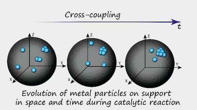
Graphical abstract. Credit: Journal of the American Chemical Society (2023). DOI: 10.1021/jacs.3c00645
A small team of chemists at the Russian Academy of Sciences, has found that metal atoms, not nanoparticles, play the key role in catalysts used in fine organic synthesis. In the study, reported in the Journal of the American Chemical Society, the group used multiple types of electron microscopy to track a region of a catalyst during a reaction to learn more about how it was proceeding.
Prior research has shown that there are two main methods for studying a reaction. The first is the most basic: As ingredients are added, the reaction is simply observed and/or measured. This can be facilitated through use of high-speed cameras. This approach will not work with nanoscale reactions, of course. In such cases, chemists use a second method: They attempt to capture the state of all the components before and after the reaction and then compare them to learn more about what happened.
This second approach leaves much to be desired, however, as there is no way to prove that the objects under study correspond with one another. In recent years, chemists have been working on a new approach: Following the action of a single particle during the reaction. This new method has proven to have merit but it has limitations as well—it also cannot be used for reactions that occur in the nanoworld. In this new effort, the researchers used multiple types of electron microscopy coupled with machine-learning algorithms.
To test their ideas, the researchers used a carbon substrate with embedded palladium nanoparticles as a catalyst. By studying reactions using such a catalyst with several types of electron microscopes and then training machine learning algorithm with the results, they were able to track a region of the catalyst as it moved through a reaction. They were able to see that there were individual metal atoms as well as clusters in addition to the nanoparticles playing a role in the reaction. Further study showed that approximately 99% of catalytic activity was due to the palladium atoms, rather than the nanoparticles despite them making up just 1% of the palladium mass.
More information: Alexey S. Galushko et al, Time-Resolved Formation and Operation Maps of Pd Catalysts Suggest a Key Role of Single Atom Centers in Cross-Coupling, Journal of the American Chemical Society (2023). DOI: 10.1021/jacs.3c00645
An artistic depiction of computer-aided structure-based rational design. The green arrows symbolize the innovative strategy to introduce new chemical properties in the hyaluronic acid molecule (green and red sticks) to develop Rationally Engineered GAG (REGAG) molecules that act as hijackers of bone regeneration blocking proteins. Dickkopf-1, a protein blocking bone formation, is shown in grey, and its receptor in beige. The colored spheres represent a set of properties that mimic the interactions between dickkopf-1 and its receptor. Credit: Gloria Ruiz Gómez
People’s ability to regenerate bones declines with age and is further decreased by diseases such as osteoporosis. To help the aging population, researchers are looking for new therapies that improve bone regeneration.
Now, an interdisciplinary team of researchers from the Biotechnology Center (BIOTEC) and the Medical Faculty of TU Dresden along with a group from Max Bergmann Center of Biomaterials (MBC) developed novel bio-inspired molecules that enhance bone regeneration in mice. The results were published in the journal Biomaterials.
As people age, their ability to regenerate bones decreases. Fractures take longer to heal and diseases like osteoporosis only add to it. This represents a serious health challenge to the aging population and an increasing socioeconomic burden for the society. To help combat this issue, researchers are looking for new therapeutic approaches that can improve bone regeneration.
A team of scientists from Dresden used computer modeling and simulations to design novel bio-inspired molecules to enhance bone regeneration in mice. The new molecules can be incorporated into biomaterials and applied locally to bone defects. These new molecules are based on glycosaminoglycans, which are long-chained sugars such as hyaluronic acid or heparin.
A sweet solution for an old bone
“Thanks to our group’s work and the work of other researchers, we know a distinct molecular pathway that regulates bone formation and repair. In fact, we can narrow it down to two proteins that work together to block bone regeneration, sclerostin and dickkopf-1” explains Prof. Lorenz Hofbauer, “The big challenge for developing drugs that improve bone healing is to efficiently turn off both of these proteins, which act as brake signals, at the same time.”
An interdisciplinary approach was a key to this challenge. The Structural Bioinformatics group led by Prof. Maria Teresa Pisabarro at the Biotechnology Center (BIOTEC) of TU Dresden and the Functional Biomaterials group led by PD Dr. Vera Hintze at the Max Bergmann Center of Biomaterials (MBC), Institute of Materials Science of TU Dresden combined their know-how with bone expert Prof. Lorenz Hofbauer at the Medical Faculty of TU Dresden.
“For several years, we have harnessed the power of computer simulations to investigate how proteins regulating bone formation interact with their receptors. All this to design new molecules that can efficiently interfere with these interactions. We worked in tandem between the computer and the bench, designing new molecules and testing them, feeding the results back to our molecular models and learning more about the molecular properties required for our goal,” explains Prof. Pisabarro.
Finally, the team of Lorenz Hofbauer’s Bone Lab used a biomaterial loaded with the new molecules on bone defects in mice to test their effectiveness. The group found that materials containing the novel molecules outperformed the standard biomaterial and enhanced bone healing by up to 50%, which indicates their potential for improving bone regeneration.
Value-added chain: From computer to the lab bench and back
The multidisciplinary team used rational drug design to create novel molecules with tailored properties and minimal side effects. By using computational methods to predict and refine the properties of the designed molecules, the team was able to develop a series of candidates with the greatest potential for turning off the proteins that block bone regeneration.
Pisabarro group’s expertise allowed the thorough analysis of the three-dimensional (3D) structures of the two proteins that block bone regeneration. With that, they were able to model their interaction with their receptors in 3D and identify so-called hot spots, i.e., specific physicochemical and dynamic properties that are essential for the biological interaction to occur.
“We used molecular modeling to design new structures that mimic relevant receptor interactions with both proteins. We wanted this binding to be stronger than their natural interactions. In this way, our novel molecules would simultaneously hijack the proteins and effectively turn them off to turn the bone regeneration on,” explains Prof. Pisabarro.
“The molecules designed by Pisabarro’s group were synthesized by our colleagues at the Free University of Berlin and then analyzed regarding their protein binding properties via biophysical interaction analysis,” says PD Dr. Hintze. “For each of the molecules we were able to measure the binding strength with the proteins and their interference with natural receptor binding of the proteins. Thus, we could reveal empirically how effective each of the small molecules could be at turning off the inhibitory proteins.” Hofbauer group then tested the biological relevance of these interaction studies in a cell culture model and later in mice.
The results of such iterative testing are a valuable asset that enhances the current molecular models of the Pisabarro group and can be used to guide the development of novel and better molecules in the future. Such an approach also ensures that animal research is minimized and enters the project only in its final phase.
The team’s findings represent an exciting step forward in preclinical development. The newly designed molecules could potentially be used to turn off the proteins that block bone regeneration and lead to the development of novel, more effective treatments for bone fractures and other bone-related conditions.
More information: Gloria Ruiz-Gómez et al, Rational engineering of glycosaminoglycan-based Dickkopf-1 scavengers to improve bone regeneration, Biomaterials (2023). DOI: 10.1016/j.biomaterials.2023.122105
Schematic illustrations showing existing and proposed ion-selective polymer membranes with varying ion channels. a, Membranes with flexible ion channels. These contain microphase-separated morphology derived from the assembly of hydrophilic ion-conductive moieties and hydrophobic flexible-polymer backbones, represented by Nafion. b, Ion-selective microporous membranes with semirigid ion channels. The channels are formed by intrinsic micropores resulting from inefficient polymer chain packing, represented by polymers of intrinsic microporosity and their derivatives. To render the membrane ion conductive, functional moieties are incorporated during postsynthetic modification. Membranes may age over time and swell in water. c,d, Our proposed membranes with rigid ion channels (c). These are expected to build from bottom-up synthesis and via swelling-resistant 3D polymer frameworks (d). Pore architecture and chemistry are tuned for rapid and selective ion transport. e,f, Preparation of stand-alone CTF membranes via a superacid-catalyzed organic sol-gel reaction from functional aromatic nitrile monomers (e). CTF membranes have a controlled number of ion-conductive moieties inside membrane pores and a covalent network structure. Image (f) shows a free-standing CTF membrane with a diameter of over 10 cm. Structure rigidity and microporosity of the CTF membrane can be regulated by designing variable structural units, as demonstrated at bottom right, from flexible to very rigid. Credit: Nature (2023). DOI: 10.1038/s41586-023-05888-x
Ion-transport membranes are vital components of clean-energy technologies, such as CO2 electrolyzers, water electrolyzers, fuel cells, redox flow batteries and ion-capture electrodialysis. These membranes must screen out specific substances to prevent crossover while efficiently conducting specific ions.
Polymer materials have the advantages of low cost, manufacturing scalability and small footprint, and thus dominate the use of ion-transport membranes in practical modules. However, the existing polymer membranes suffer from a ubiquitous “conductivity-selectivity” trade-off: highly conductive membranes tend to exhibit low selectivity and vice versa. This trade-off presents a challenge in developing membrane materials that meet the required performance criteria.
In a study published in Nature on April 26, the research team led by Professor Xu Tongwen and Professor Yang Zhengjin from the University of Science and Technology of China (USTC) of the Chinese Academy of Sciences (CAS), and their collaborators, proposed a new type of ion exchange membrane—triazine framework polymer membranes—which can break the conductivity-selectivity trade-off.
Compared to traditional materials, the triazine framework polymer membranes exhibited much enhanced capacity in both anti-swelling and anti-aging, showing an extremely low swelling ratio on water absorption. Their rigid channels ensured high selectivity from size-sieving, thus enabling extremely low permeability of active materials.
With proper control over the chemistry of rigid pore channels, the researchers observed near-frictionless ion flow within the all-rigid triazine framework polymer membrane (SCTF-BP), with the ion diffusion coefficient close to value in water. This is achieved by the robust micropore confinement within the rigid pore channels and multi-interaction between ion and membrane.
These framework membranes exhibited both extremely low permeability of active materials and ultrahigh ion diffusivity, and their advantages were exemplified as ion-conducting membranes in 2,6-dihydroxy anthraquinone / K4[Fe(CN)6] aqueous organic redox flow batteries. The membrane delivered a neat area-specific resistance as low as 0.17 Ω cm2, and thus enabled stable cell operation at extreme current densities, from 200 to 500 mA cm-2, with both high energy efficiency and high-capacity utilization.
These data related to energy efficiency and capacity utilization far surpass those for otherwise identical cells assembled with commercial membranes and state-of-the-art ion-sieving membranes.
This work highlights the importance of secondary interactions to develop high-performing ion-transport membranes. The design strategy proposed is believed to be broadly applicable, considering numerous options of organic reactions and functional monomers that can be utilized to construct polymer frameworks, and directs the fit-for-purpose design of membranes according to practical application demand.
More information: Peipei Zuo et al, Near-frictionless ion transport within triazine framework membranes, Nature (2023). DOI: 10.1038/s41586-023-05888-x
The human body relies heavily on electrical charges. Lightning-like pulses of energy fly through the brain and nerves and most biological processes depend on electrical ions traveling across the membranes of each cell in our body.
These electrical signals are possible, in part, because of an imbalance in electrical charges that exists on either side of a cellular membrane. Until recently, researchers believed the membrane was an essential component to creating this imbalance. But that thought was turned on its head when researchers at Stanford University discovered that similar imbalanced electrical charges can exist between microdroplets of water and air.
Now, researchers at Duke University have discovered that these types of electric fields also exist within and around another type of cellular structure called biological condensates. Like oil droplets floating in water, these structures exist because of differences in density. They form compartments inside the cell without needing the physical boundary of a membrane.
Inspired by previous research demonstrating that microdroplets of water interacting with air or solid surfaces create tiny electrical imbalances, the researchers decided to see if the same was true for small biological condensates. They also wanted to see if these imbalances sparked reactive oxygen, “redox,” reactions like these other systems.
Appearing on April 28 in the journal Chem, their foundational discovery could change the way researchers think about biological chemistry. It could also provide a clue as to how the first life on Earth harnessed the energy needed to arise.
“In a prebiotic environment without enzymes to catalyze reactions, where would the energy come from?” asked Yifan Dai, a Duke postdoctoral researcher working in the laboratory of Ashutosh Chilkoti, the Alan L. Kaganov Distinguished Professor of Biomedical Engineering and Lingchong You, the James L. Meriam Distinguished Professor of Biomedical Engineering.
“This discovery provides a plausible explanation of where the reaction energy could have come from, just as the potential energy that is imparted on a point charge placed in an electric field,” Dai said.
When electric charges jump between one material and another, they can produce molecular fragments that can pair up and form hydroxyl radicals, which have the chemical formula OH. These can then pair again to form hydrogen peroxide (H2O2) in tiny but detectable amounts.
“But interfaces have seldom been studied in biological regimes other than the cellular membrane, which is one of the most essential part of biology,” said Dai. “So we were wondering what might be happening at the interface of biological condensates, that is, if it is an asymmetric system too.”
Cells can build biological condensates to either separate or trap together certain proteins and molecules, either hindering or promoting their activity. Researchers are just beginning to understand how condensates work and what they could be used for.
Because the Chilkoti laboratory specializes in creating synthetic versions of naturally occurring biological condensates, the researchers were easily able to create a test bed for their theory. After combining the right formula of building blocks to create minuscule condensates, with help from postdoctoral scholar Marco Messina in? Christopher J. Chang’s group at the University of California—Berkeley, they added a dye to the system that glows in the presence of reactive oxygen species.
Their hunch was right. When the environmental conditions were right, a solid glow started from the edges of the condensates, confirming that a previously unknown phenomenon was at work. Dai next talked with Richard Zare, the Marguerite Blake Wilbur Professor of Chemistry at Stanford, whose group established the electric behavior of water droplets. Zare was excited to hear about the new behavior in biological systems, and started to work with the group on the underlying mechanism.
“Inspired by previous work on water droplets, my graduate student, Christian Chamberlayne, and I thought that the same physical principles might apply and promote redox chemistry, such as the formation of hydrogen peroxide molecules,” Zare said. “These findings suggest why condensates are so important in the functioning of cells.”
“Most previous work on biomolecular condensates has focused on their innards,” Chilkoti said. “Yifan’s discovery that biomolecular condensates appear to be universally redox-active suggests that condensates did not simply evolve to carry out specific biological functions as is commonly understood, but that they are also endowed with a critical chemical function that is essential to cells.”
While the biological implications of this ongoing reaction within our cells is not known, Dai points to a prebiotic example of how powerful its effects might be. The powerhouses of our cells, called mitochondria, create energy for all of our life’s functions through the same basic chemical process. But before mitochondria or even the simplest of cells existed, something had to provide energy for the very first of life’s functions to begin working.
Researchers have proposed that the energy was provided by thermal vents in the oceans or hot springs. Others have suggested this same redox reaction that occurs in water microdroplets was created by the spray of ocean waves.
But why not condensates instead?
“Magic can happen when substances get tiny and the interfacial volume becomes enormous compared to its volume,” Dai said. “I think the implications are important to many different fields.”
More information: Yifan Dai et al, Interface of biomolecular condensates modulates redox reactions, Chem (2023). DOI: 10.1016/j.chempr.2023.04.001
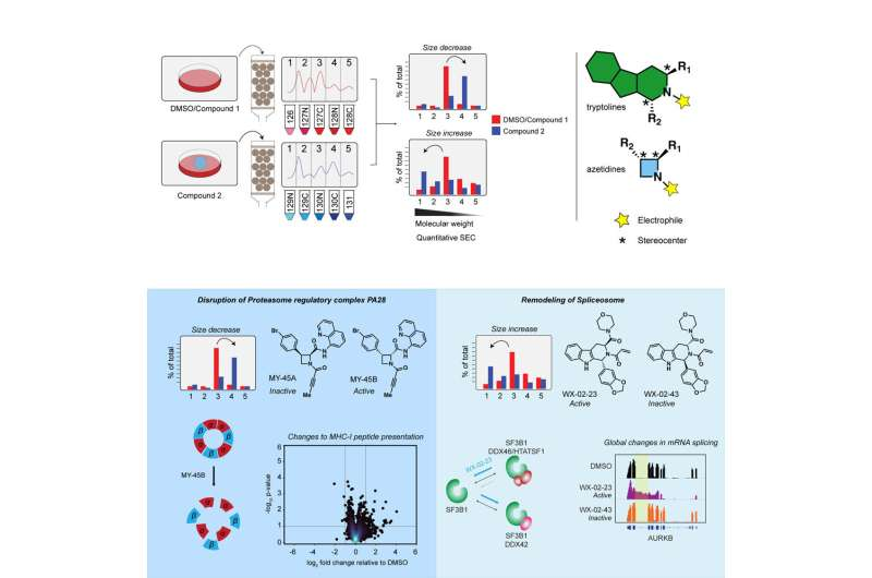
Scripps Research scientists have developed a new strategy for identifying small molecules that can change the function of proteins, offering a promising path for discovering targeted drugs. In collaboration with scientists at other institutions, the group used their new approach to find small molecules that can alter the activity of proteins involved in cancer.
The research, published in Molecular Cell on April 20, improves on previous methods that could screen for whether small molecules selectively attached to proteins, but not whether they affected the proteins’ biological activities. The new method revolves around using two mirror-image versions of a small molecule and comparing how they change the size of protein complexes in cells.
“The ability of small molecules to specifically bind to a protein and cause a biological consequence is the fundamental basis for most drugs today,” says senior author Benjamin Cravatt, Ph.D., Gilula Chair of Chemical Biology at Scripps Research. “With this assay, we’re expanding our ability to discover these small molecules that not only bind proteins, but have functional impacts.”
In recent years, Cravatt’s lab has designed sets of small chemicals that can irreversibly bind to certain parts of proteins. However, screening these chemical libraries to discover their possible impact on protein function was generally a slow and tedious process. Since individual proteins have different roles in cell biology, researchers often have to develop specialized functional screens for each protein of interest. One screen, for instance, might determine whether the chemicals affected cell growth, while another might determine whether the chemicals changed levels of a different molecule.
“Just because a small molecule engages a protein physically doesn’t mean that it changes the protein’s function in the cell,” says co-first author Jarrett Remsberg, Ph.D., who carried out the work as an American Cancer Society postdoctoral research fellow in the Cravatt lab at Scripps Research. Former graduate student Michael Lazear, Ph.D. and postdoctoral fellow Martin Jaeger, Ph.D. were also first authors of the paper.
In the new work, Cravatt’s group used the conglomeration of proteins into complexes as a proxy for their function. Proteins often work by binding to other proteins—if this binding doesn’t happen or if it is induced to happen, it indicates a protein’s function may have changed.
The research team designed pairs of “mirror image” molecules, called stereoisomers, that could each bind irreversibly to proteins in the same way that their previous chemical libraries had worked. The pairs of stereoisomers let them be sure that the impact of each small molecule was due to its unique structure (if only one version of a molecule changes the proteins’ function, it is likely a specific and direct interaction).
Once they exposed cells to the pairs of stereoisomers, they tested whether a protein-of-interest was in a different size complex, using a technique called size exclusion chromatography in which proteins are sifted through beads with different sized pores.
To show the utility of this approach, the researchers screened the set of small molecules for their ability to change the sizes of protein complexes in prostate cancer cells. They pinpointed a molecule, MY-1B, which selectively disrupted a complex of proteins known as PA28, previously found to play a role in degrading proteins in cancer. Further work in leukemia cells confirmed that, by specifically binding to the protein PMSE1, MY-1B or a related compound (but not their mirror images) could effectively inactivate the PA28 complex.
Cravatt and colleagues also followed up on an observation that a different chemical, EV-96, changed the size of a protein complex involved in splicing strands of RNA inside cells. The team discovered that EV-96 slowed the growth of cancer cells and pinpointed SF3B1 as the protein the chemical was binding to.
In both cases, the new chemicals represent the first time scientists have been able to target the protein complexes—PA28 and the so-called spliceosome— with small, simple synthetic chemicals.
“This means that researchers have new chemical tools in their arsenal that they didn’t have before,” says Remsberg. “It’s an opportunity for better understanding these proteins as well as investigating potential therapeutic opportunities.”
The team hopes their approach can be expanded to use other functional readouts than complex size, and they intend to use it to study different cell types in the future.
“The long-term idea is that we can use this approach to discover chemical compounds that impinge upon any readout,” says Cravatt. “There are certainly other readouts that we hope to be able to look at in the future.”
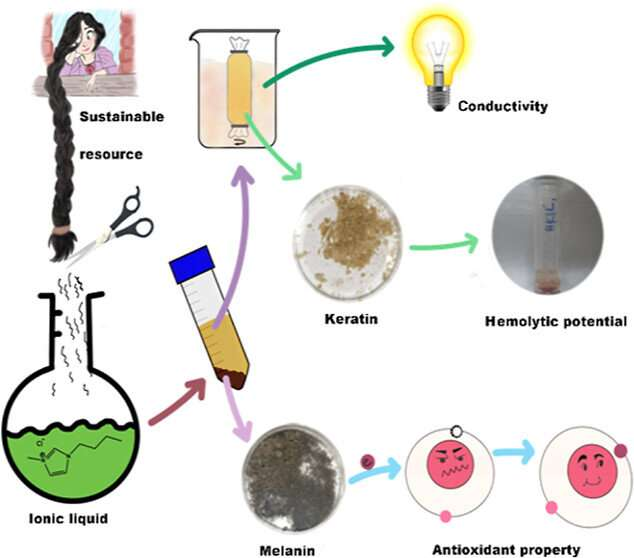
Hair styling can be a potent form of self-expression, whether it features dramatic updos, intricate braids or crazy colors. Beyond being a reflection of our personality, these strands contain compounds that could one day appear in bandages, sunscreens or other products. Researchers reporting in ACS Omega have now designed a simple, green process to extract both keratin and melanin from human hair for these possible applications without harsh chemicals or excessive waste.
Hair is made up of protein filaments consisting of many different layers and components. Its structure comes from the protein keratin, which can also be found in fingernails, horns and feathers. Its color is provided by melanin, a group of pigment molecules that are also found in the skin and eyes. In addition, melanin has antioxidative properties and can help shield against ultraviolet light.
These qualities make the compounds suitable for biomedical applications; however, since most discarded hair is incinerated or dumped in landfills, its keratin and melanin are largely unused as well. Chemically extracting them from hair is possible, but current protocols either can only extract one compound at a time, or rely on harsh chemicals and complicated steps. So, Paulomi Ghosh and colleagues wanted to develop a straightforward method to extract both keratin and melanin from human hair with a single procedure, using a recyclable, green solvent.
The researchers collected samples of hair from local salons, then washed and cut them into small slices. Then, they mixed the hair with an ionic liquid, which dissolved the mixture by interrupting the hydrogen bonds that held the keratin proteins together. When heated and poured into a hydrochloric acid solution, the melanin pigments precipitated out and were collected. Next, the researchers performed dialysis to collect the keratin proteins. The ionic liquid was recycled and reused in subsequent reactions, without a significant impact on the reaction’s yield.
Recovered keratin was compatible with blood, suggesting that it could be used in heavy-duty hemostatic bandages. This extraction procedure also maintained the natural structure of the melanin, which was lost in other, harsher methods. Because the melanin had good antioxidative and UV shielding properties, the team says it could be used in sun-protective products or films. The researchers say that this technique could serve as a green way to sustainably extract useful biopolymers from otherwise discarded materials.
More information: Ashmita Mukherjee et al, One-Pot Extraction of Bioresources from Human Hair via a Zero-Waste Green Route, ACS Omega (2023). DOI: 10.1021/acsomega.3c01428
Cryo-EM formed the basis for deciphering the structure of a receptor involved in many bodily processes. Credit: Kruse lab
As the proverb goes, it takes a village to raise a child. It can also take a village to make progress in science and medicine.
In this case, labs from not one, not two, but three departments in the Blavatnik Institute at Harvard Medical School, along with a colleague in France, came together to figure out the structure of a tricky receptor involved in heart, lung, liver, and kidney function as well as pregnancy.
Untangling this structure, described April 20 in Nature Chemical Biology, provides a foundation for developing drugs that act on the receptor with the goal of treating heart disease and conditions marked by the buildup of scar tissue, or fibrosis. Those include idiopathic pulmonary fibrosis, a chronic disease of the lungs; non-alcoholic fatty liver disease; and scleroderma, which affects the skin, joints, and internal organs.
The work may also pique the interest of other scientists who study basic biology because the receptor has an unusual structure that stands out from other members of its family, the G protein-coupled receptors.
“This was a very difficult receptor to characterize,” said senior author Andrew Kruse, HMS professor of biological chemistry and molecular pharmacology. “The collaboration of multiple labs was essential, and the project highlights the value of combining experimental and computational methods.”
“You have a lot of great opportunities at HMS to work with talented scientists who use different techniques than you,” agreed Sarah Erlandson, who led the work as a graduate student in the Kruse lab. “That allowed us to form a more complete picture of the receptor and study it in more ways than we could by ourselves.”
A relaxin possibility
The receptor’s name is a mouthful: relaxin/insulin-like family peptide receptor 1, or RXFP1. It gets that name because it and its three receptor siblings bind to hormones called relaxins.
Relaxins are best known for initiating a constellation of changes in the body during pregnancy, including relaxing ligaments in the pelvis in preparation for childbirth. It also boosts sperm movement. But scientists have grown to appreciate the hormone’s many non-reproductive roles as well, including dilating blood vessels to increase blood flow, stimulating the growth of new blood vessels, breaking down collagen, and reducing inflammation and fibrosis.
Relaxins do all of this when they get released from tissues such as the heart, prostate, and placenta and bind to receptors in the membranes of cells in certain organs. The receptors then send signals that spur the cells to act.
Most relaxins stay local, but one type, relaxin-2, travels throughout the body via the blood. This is the relaxin that binds to RXFP1.
Relaxin-2’s involvement in so many bodily processes has turned scientists’ eyes toward treating diseases by mimicking higher or lower levels of the hormone. Doing so requires designing drugs that bind to RXFP1—which is hard to do without knowing its structure.
“There are no drugs available that target this receptor,” said Erlandson, who now works as a research scientist at Takeda Pharmaceuticals. “People are interested in it as an option for treatment of cardiovascular and fibrotic diseases, but when you don’t understand the detailed structure, it limits your ability to target it.”
So, the teams got to work.
It takes two to tango, four to solve a receptor structure
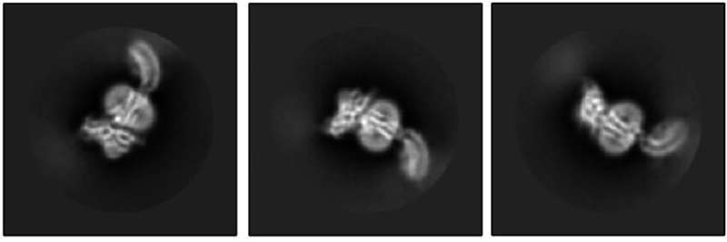
The Kruse lab took the first step by using cryo-electron microscopy to reveal what RXFP1 looks like at the near-atomic level when bound to relaxin-2.
But one blurry spot remained where a flexible part of the receptor kept changing position from one snapshot to the next. That elusive part was the most important one—the part that binds to relaxin.
Members of the lab of Steven Gygi, HMS professor of cell biology, tackled the problem using mass spectrometry, a different method for determining structural information that measures atomic weights. Combining the experimental cryo-EM results with the mass spectrometry data allowed Erlandson to fill in the missing structural details.
Now the researchers could see the receptor in its active, or “on,” state, bound to relaxin-2. The structure suggested that the receptor could turn itself on. If that were true, what prevented it from being on all the time, sending cell-activation signals whether relaxin was there or not?
Insight came from Debora Marks, HMS associate professor of systems biology, and Xiaojing Cong at the Institute of Functional Genomics (IGF) in France. They used computational techniques—including one called evolutionary coupling analysis, which looks at protein sequences that change together over time—to predict how different parts of the receptor might shift around between its active and inactive states.
At last, the story revealed itself.
When RXFP1 is alone, no relaxins in sight, it’s turned off. When relaxin-2 binds to it, multiple parts of the receptor change shape and communicate with one another to flip the “on” switch.
The collaboration allowed the team to answer open questions about how RXFP1’s multiple parts move around and work together to allow the receptor to do its job. The way it binds to relaxin-2 hasn’t been seen in many of its receptor relatives.
With RXFP1’s active structure in hand, researchers now have a lock to design therapeutic keys for.
“It was really exciting to get to the point in the project where we saw the structure and were making these discoveries,” said Erlandson. “There’s more work to be done, but this makes a big contribution that benefits scientists and could ultimately help patients.”
Additional authors are Shaun Rawson, James Osei-Owusu, Kelly P. Brock, Xinyue Liu, Joao A. Paulo, and Julian Mintseris, of HMS.
More information: Sarah C. Erlandson et al, The relaxin receptor RXFP1 signals through a mechanism of autoinhibition, Nature Chemical Biology (2023). DOI: 10.1038/s41589-023-01321-6
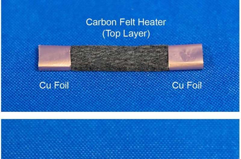
The assembly of the STH system. A thinner layer of carbon felt (about 2.3 mm) is used as the top heater layer. The two ends of the top heater layer are wrapped with Cu foil electrodes for Joule heating. The top and bottom layers are placed in soft contact (without external pressure) for the STH process. A quartz tube with 10.5 mm inner diameter was used to contain the carbon bilayer structure and the reactant reservoir. The bottom image shows the heater layer exhibiting a bright orange color as we apply an electrical current through the top heater layer, demonstrating its Joule-heating capabilities. Credit: Nature (2023). DOI: 10.1038/s41586-023-05845-8
A team of engineers and materials scientists affiliated with multiple institutions in the U.S., has developed a new way to depolymerize plastics using electrified spatiotemporal heating. In their paper, published in the journal Nature, the group describes the new process and its efficiency. Nature has also published a Research Briefing in the same journal issue outlining the work done by the team.
Over the past several years, plastic pollution has become a major concern, both for the environment and for the health of plants and animals, including humans, and scientists are seeking ways to recycle it. Most of the techniques developed thus far involve using chemicals to depolymerize plastics. These efforts are still extremely inefficient, however, with yields between 10% and 25%. In this new effort, the team has found a way to use pulsed electricity to boost the yield to approximately 36%.
The approach involved designing a new kind of reactor with a porous carbon felt bilayer and a pulsed electric heater at the top. In their reactor, plastic bits are melted as they are fed in to the upper chamber and flow as a mass into a lower chamber, where the material is pushed through the felt filter. The plastic then begins to decompose as the temperature rises. As the molecules that make up the plastic become smaller, their volatility grows until they are expelled from the reactor as a gas, which allows more liquid to be drawn in. Using electricity to heat the plastic allows for oscillating the temperature, allowing simpler depolymerization reactions to take precedence over side reactions, which need additional heating to depolymerize.
In addition to improving efficiency, the new approach uses less energy because of the oscillating instead of constant heat source. The team notes the system could be made more eco-friendly by using renewable sources for the electricity. They note that their reactor does emit other materials, such as acetylene, methane and some larger molecules, along with some aromatics. They also acknowledge that more work is required to reduce the amount of carbon released during the reactions.
More information: Qi Dong et al, Depolymerization of plastics by means of electrified spatiotemporal heating, Nature (2023). DOI: 10.1038/s41586-023-05845-8
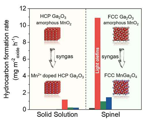
Crystal-phase-dependent of syngas conversion activity of MnGaOx-SAPO-18. Credit: Bai Bing and Jiao Feng
Two notable metal oxide structures, spinel-type oxide and solid solution-type oxide, are widely used in Oxide-Zeolite (OXZEO) bifunctional catalysts for CO/CO2 hydrogenation reactions.
Identifying the crystallographic structure sensitivity of catalysts in chemical reactions is helpful to the rational design of catalysts. However, direct and convincing study to correlate the crystal structure of oxide to its catalytic performance has yet to be done.
Recently, a joint research team led by Prof. Bao Xinhe, Prof. Pan Xiulian, Assoc. Prof. Jiao Feng and Prof. Xiao Jianping from the Dalian Institute of Chemical Physics (DICP) of the Chinese Academy of Sciences (CAS) has observed strong crystal phase-dependent activity of MnGaOx in direct syngas conversion. This study was published in Angewandte Chemie International Edition on April 18.
“The main challenge to observing the crystal structure of oxide is the lack of a well-defined material-synthesis method to obtain metal oxides with the same element components but different crystallographic structures,” said Jiao.
In this study, the researchers found co-precipitation method and hydrothermal method could synthesize bimetallic oxides, which were composed of amorphous MnO2 and Ga2O3 with hexagonal close-packed (HCP) or Face centered-cubic (FCC) crystal phase, respectively. More interestingly, they found that the HCP oxide remained unchanged as HCP MnO-Ga2O3 solid solution oxides after reduction under H2 or CO, while the FCC solid solution oxide transformed into FCC spinel structure, where reduced Mn2+ took the A-site of AB2O4 spinel structure.
They obtained 40% CO conversion, 81% light olefins selectivity, and 0.17 g·gcat-1·h-1 space-time yield of light olefins with the combination of FCC MnGaOx-Spinel and SAPO-18. In comparison, they obtained a much inferior activity with solid solution MnGaOx with a similar chemical composition.
They further proved that the superior activity of MnGaOx-Spinel was attributed to its higher reducibility and the presence of coordinatively unsaturated Ga3+ site, which facilitated the dissociation of the C-O bond via a more efficient ketene-acetate pathway to light olefins.
“Our findings may further optimize metal oxides for OXZEO syngas conversion,” said Prof. Pan.
More information: Bing Bai et al, Tuning the Crystal Phase to Form MnGaOx‐Spinel for Highly Efficient Syngas to Light Olefins, Angewandte Chemie International Edition (2023). DOI: 10.1002/anie.202217701
Hydrogen cyanide (HCN) featured with high volatility and high adsorption is a common toxic and hazardous gas. Traces of HCN are also found in human exhaled breath. Unusual high HCN concentration in the breath of cystic fibrosis (CF) patients is associated with Pseudomonas aeruginosa (PA) infection. Therefore, the development of a highly sensitive online HCN measurement in exhaled breath can enable rapid screening for PA infection in CF patients.
Recently, a research group led by Prof. Li Haiyang from the Dalian Institute of Chemical Physics (DICP) of the Chinese Academy of Sciences (CAS) has developed a flow-assisted photoionization mass spectrometry method for profiling hydrogen cyanide in exhaled breath.
The study was published in Analytical Chemistry on April 4.
“HCN is easily soluble in water and highly adsorbed on device surfaces, so sensitivity and response speed are main challenges for directly measuring HCN in exhaled breath at high humidity presents,” said Prof. Chen Ping, co-corresponding author of this study.
The researchers developed a method by utilizing a self-developed atmospheric pressure negative photoionization time-of-flight mass spectrometer instrument and improved the sensitivity and time resolution of direct HCN measurements in exhaled breath. This method enabled real-time tracking of HCN concentrations in a single exhaled breath, which could provide an effective means for early screening of CF patients with PA infection.
They proposed to use helium shield gas within the mass spectrometry ionization source. This approach reduced the effect of high humidity on ionization, improved ion transport efficiency, and thus enhanced HCN detection sensitivity.
Moreover, they improved the sampling system by shortening the sampling line and adding a gas purging process to effectively reduce HCN adsorption residue and improve time resolution. This method achieved a limit of detection of 0.3 ppbv and a resolution time of 0.5 s.
By utilizing this method, the researchers tracked the changes in the single exhaled HCN profiles of volunteers before and after gargling. The resulting profiles clearly showed an early peak and a stable end-tidal plateau, representing the concentration of the oral cavity and end-tidal gas, respectively.
“The new method demonstrated good resistance to interference and high accuracy in HCN quantification. It has potential applications in the detection of PA infection in CF patients,” said Prof. Li.
More information: Yuxuan Wen et al, Online Detection of HCN in Humid Exhaled Air by Gas Flow-Assisted Negative Photoionization Mass Spectrometry, Analytical Chemistry (2023). DOI: 10.1021/acs.analchem.2c05603

. DOI: 10.1021/jacs.3c00645")
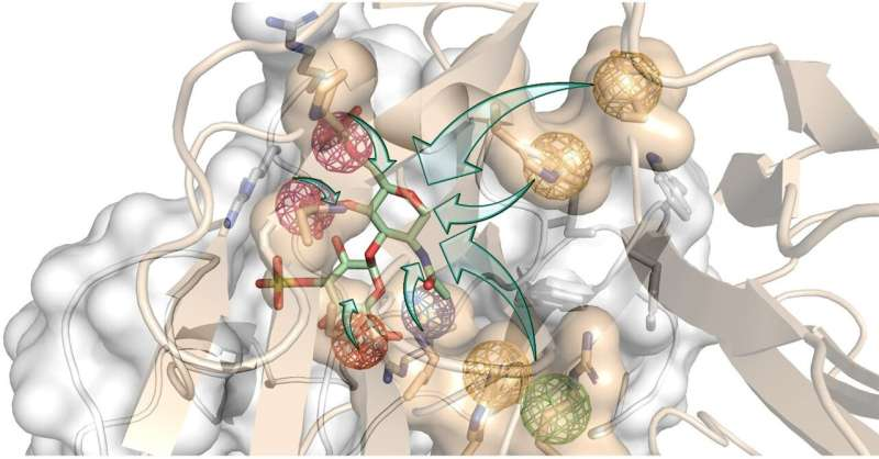
 to develop Rationally Engineered GAG (REGAG) molecules that act as hijackers of bone regeneration blocking proteins. Dickkopf-1, a protein blocking bone formation, is shown in grey, and its receptor in beige. The colored spheres represent a set of properties that mimic the interactions between dickkopf-1 and its receptor. Credit: Gloria Ruiz Gómez")
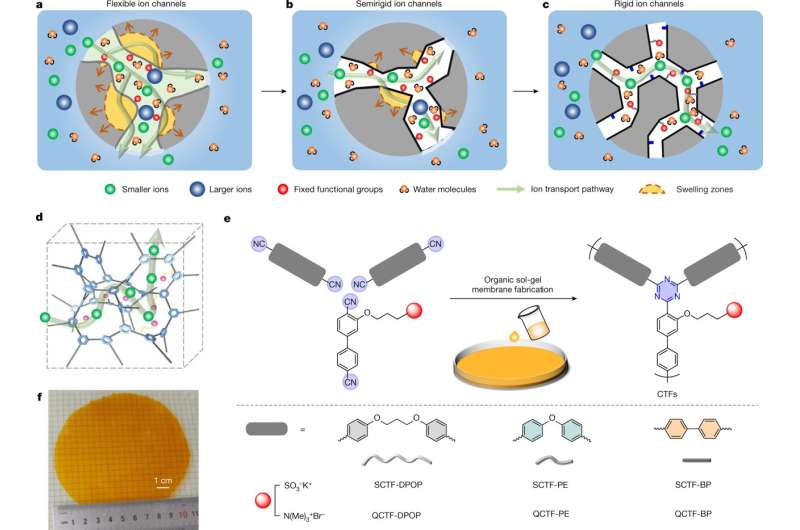
. These are expected to build from bottom-up synthesis and via swelling-resistant 3D polymer frameworks (d). Pore architecture and chemistry are tuned for rapid and selective ion transport. e,f, Preparation of stand-alone CTF membranes via a superacid-catalyzed organic sol-gel reaction from functional aromatic nitrile monomers (e). CTF membranes have a controlled number of ion-conductive moieties inside membrane pores and a covalent network structure. Image (f) shows a free-standing CTF membrane with a diameter of over 10 cm. Structure rigidity and microporosity of the CTF membrane can be regulated by designing variable structural units, as demonstrated at bottom right, from flexible to very rigid. Credit: Nature (2023). DOI: 10.1038/s41586-023-05888-x")

. DOI: 10.1016/j.molcel.2023.03.026")
. DOI: 10.1021/acsomega.3c01428")

 is used as the top heater layer. The two ends of the top heater layer are wrapped with Cu foil electrodes for Joule heating. The top and bottom layers are placed in soft contact (without external pressure) for the STH process. A quartz tube with 10.5 mm inner diameter was used to contain the carbon bilayer structure and the reactant reservoir. The bottom image shows the heater layer exhibiting a bright orange color as we apply an electrical current through the top heater layer, demonstrating its Joule-heating capabilities. Credit: Nature (2023). DOI: 10.1038/s41586-023-05845-8")


. DOI: 10.1021/acs.analchem.2c05603")