
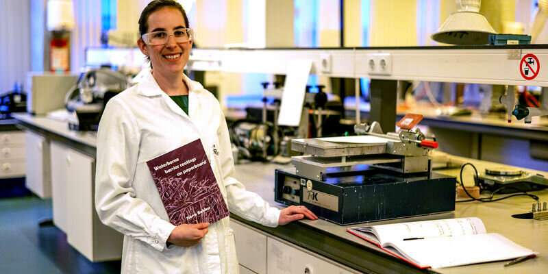

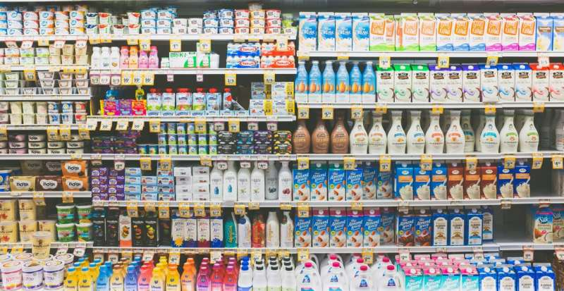
On average, we open seven packaged items per day, most of them food items. All of this together makes for a mountain of plastic. But more and more often our tomatoes, apples and cookies are packaged in cardboard. To help speed up the transition of plastic to paper, TU/e chemist Sterre Bakker researched what coatings can be used to make cardboard a more suitable food packaging material.
About 20% of waste consists of packaging, but packaging food is not all bad. Good packaging protects the item during transport, which means less food needs to be thrown out. Packaging also keeps out moisture, bacteria and fungi. This gives our food a longer shelf life, also resulting in less food going to waste. Keeping a bag of mixed vegetables or a pack of beef in the fridge for a week without it going off? Impossible without the packaging.
More alternatives are being used to reduce plastic packaging waste. Over the past few years, paper and cardboard have become more common packaging materials. But transitioning from plastic to paper is not as easy as it seems, TU/e researcher Sterre Bakker explains. “Plastic has a number of highly practical characteristics. You can use it for airtight packaging and it is an excellent barrier for water and grease. It’s strong but also light, which works well for the transport sector. It requires a lot of effort to find a suitable alternative that meets the same requirements and can be used at a large scale.”
Freedom to publish
For her Ph.D., Bakker investigated how cardboard can be used to make food packaging more sustainable. This Friday (Jan. 27, 2023) she will defend her thesis at the department of Chemical Engineering and Chemistry. To bridge the gap between the laboratory and the packaging sector, she collaborated with chemicals company BASF.
When asked if this collaboration caused any confidentiality issues, Bakker shakes her head. “I consciously sought collaboration with industry; I wanted to get a PHD, but in applied research. This allowed me to make a contribution to society. As I wanted to have the freedom to talk and publish about my findings, we used a model coating. It’s not exactly the same as the manufacturer’s, but it’s very similar. So that was a good compromise.”
Water-based coating
For dry food items, such as rice and oatmeal, cardboard packaging works fine. But putting milk or a hamburger in a cardboard box is more complex, Bakker explains. “A suitable coating is crucial. Water and grease have very different ways of getting through a coating. For a good water barrier, the chemical reaction that occurs while the coating is drying up is very important, but for grease you need a coating that’s intact. This explains why the inner lining of a milk carton is so thick: it consists of several layers of hydrophilic and hydrophobic coatings. In addition to the relatively large amount of plastic used, the mixture of coatings makes it hard to recycle. This is what we would like to improve.”
Bakker set out in search of a single coating impermeant to water, oxygen and grease. To this end, she studied an innovative, water-based coating, with the special addition of a water-soluble, synthetic resin to stabilize polymer particles. This resin makes it possible to transform a water-soluble surface into a water-resistant one. “When the coating dries up, it’s not only the water that evaporates but also a base. The resin undergoes a chemical reaction and this means the coating no longer dissolves in water.”
Multifunctional coating
Bakker used a scanning electron microscope and infrared spectroscopy to carry out in-depth research into the different barrier characteristics of the coating, as well as to optimize the production process. This is all very relevant to industry, she emphasizes, as synthesizing the coating is easy to scale and not overly complex. What’s more, at a certain temperature the color of the coating can be changed by adding a specific molecule. This quasi-alternative expiration date makes the coating multifunctional.
Bakker shows her final result: a shiny piece of thick carton that actually resists both water and grease. “This coating already has a lot of advantages in comparison to current coatings. Although it does require raw fossil materials, the layer can be made much thinner now. We’ve also demonstrated through several experiments that the coating can be peeled off the cardboard more easily, which means it’s very suitable for recycling. We’ve taken the first steps, obviously in the hope of eventually creating a biobased coating that eliminates all plastic from food packaging.”
Tea lover
The above should serve as a reminder, Bakker reiterates, of cardboard being much stronger than we give it credit for. Like women, she says with a smile. Bakker is quite comfortable in the space she created for herself in the male-dominated coating and packaging industry. And as a tea lover she will obviously bring her tea box—cardboard of course—to her new place of work, Allnex in Bergen op Zoom. Before we leave she’s keen to show us the quote adorning her thesis: “A woman is like a teabag; you never know how strong it is until it is in hot water.”
Sterre Bakker defends her thesis at the department of Chemical Engineering and Chemistry on January 27, 2023.
Provided by Eindhoven University of Technology


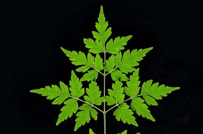
, a mahogany species. Credit: John Innes Centre")
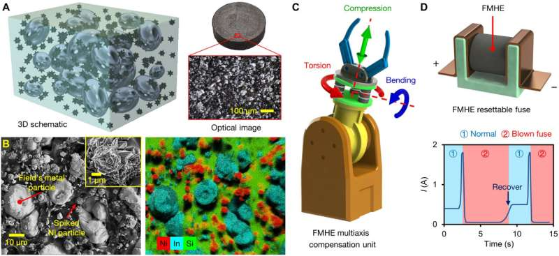
 3D schematic of the composition and optical microscope image showing the surface structure of the FMHE. ( B ) SEM and EDS images of the cross section of the FMHE 3 sample. The distribution of Ni and FM particles; PDMS are represented by their characteristic elements of Ni, In, and Si. The inset shows a close-up SEM image of a spiked Ni particle. ( C ) An FMHE variable stiffness compensation unit (highlighted in green) mounted on a robot manipulator. ( D ) Schematic diagram of the FMHE resettable current-liming fuse and its current-time curve during cyclic operation. Credit: Science Advances (2023). DOI: 10.1126/sciadv.adf1141")
. DOI: 10.1021/acsami.2c14872")
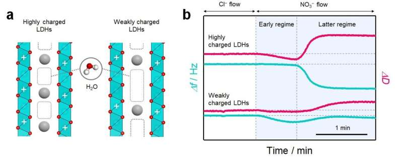
 Schematic of the interlayer structure in layered materials with different host charge densities. In the interlayer space, water molecules are incorporated into the pores that are not filled with charge compensating ions for host charge. (b) Quartz crystal microbalance with energy dissipation monitoring (QCM-D) profiles of ion-exchange reaction in LDHs with different host charge densities showing the change in the frequency (Δf) and dissipation (ΔD). Credit: Nature Communications (2022). DOI: 10.1038/s41467-022-34124-9")
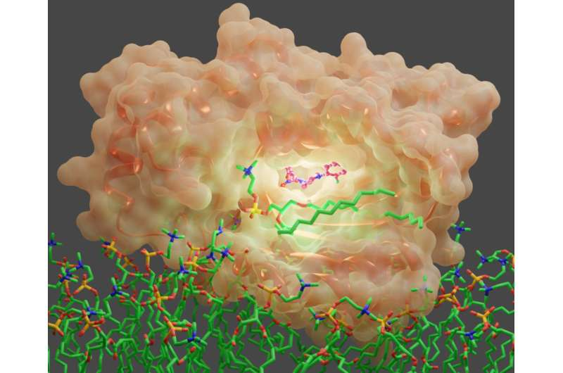
, a potential therapeutic target in drug-resistant fungal infections, in complex with a small molecule inhibitor (purple). Credit: Sachin S. Katti")


")
