What do oranges, coffee grounds and seaweed have in common? They outshine cotton in sustainable fashion

What do oranges, coffee grounds and seaweed have in common? They outshine cotton in sustainable fashion
Credit: Shutterstock

Ever considered the carbon footprint of manufacturing your favorite shirt?

The average cotton shirt produces 2.1 kilograms of carbon dioxide—but a polyester shirt produces over twice as much (5.5 kilograms). It might come as no surprise that the fashion industry is responsible for around 5% of global CO₂ emissions.

Some natural fibers can also take a heavy toll on the environment. Last week, for example, an ABC investigation revealed hundreds of hectares of the Northern Territory’s pristine tropical savanna had been cleared to make way for cotton farms, sometimes without permit.

So are there more sustainable textiles we should be producing and purchasing instead?

Research, including our own ongoing research, points to certain “non-traditional fibers” as new green alternatives. These include fibers produced from wastes—think coffee waste and recycled plastic bottles—as well as seaweed, orange, lotus, corn and mushroom.

Brands such as PatagoniaMud JeansNinety PercentPlant Faced Clothing and Afends are among the brands leading the way in incorporating sustainable fibers into their products. But the true turning point will likely come when more of the biggest names in fashion get involved, and it’s high time they invest.

The problems with traditional fibers

There are two types of traditional fibers: natural and synthetic. Natural fibers, such as cotton and flax, have certain advantages over synthetic fibers which are derived from oil and gas.

When sustainability is considered, natural fibers are preferred over the synthetic fibers due to, for instance, their ability to biodegrade and their availability in the environment.

However, some natural fibers (particularly cotton) need a lot of fresh water and chemicals that are toxic to the environment for harvesting. For example, it takes 10,000 liters of water on average to grow just 1 kilogram of cotton.

In comparison, synthetic fibers consume a significantly lower amount of water (about one hundredth), but a significantly higher amount of energy.

Petrochemical fibers made from fossil fuels—such as polyester, nylon and acrylic—are the backbone of fast fashion. Yet another big problem with such products is that they don’t easily decompose.

As they slowly break down, petrochemical fibers release microplastics. These not only contaminate the environment, but also enter the food chain and pose health risks to animals and humans.

You may have also come across blended fabrics, which are produced with a combination of two or more types of fibers. But these pose challenges in sorting and recycling, as it’s not always possible or easy to recover different fibers when they’re combined.

Non-traditional fibers: a potential game changer

Amid the overconsumption of traditional fibers, several global fashion brands have started to adopt new fibers derived from seaweed, corn, and mushroom. This includes Stella McCartneyBalenciagaPatagonia, and Algiknit.

Other emerging natural fibers include lotus, pineapple and banana fibers. Lotus fibers are extracted from the plant stem, banana fibers are extracted from the petiole (the stalk that connects the leaf and stem), and pineapple fibers are extracted from pineapple leaves.

The process of extracting fibers from wastes such as orange peelscoffee grounds, and even from the protein of waste milk, has also been well researched, and clothes have been successfully manufactured from these materials.

All these examples of non-traditional fibers are free from many of the problems mentioned earlier, such as heavy resource consumption (particularly fresh water), use of toxic chemicals, and the use of large amounts of energy (for synthetic fibers).

Further, these fibers are biodegradable at their end of life and don’t release microplastics when you wash them.

Meanwhile, there has been tremendous growth in the use of recycled synthetic fibers, which reduces the use of virgin materials, energy and chemical consumption. Recycling plastics such as drink bottles to make clothing is also becoming more common. Such innovations can help lower our dependency on raw materials and mitigate plastic pollution.

What’s more, the selection of appropriate color combinations during recycling and processing for fabrics can avoid the need for dyeing.

What now?

Fashion companies can reduce the load on the environment through seriously investing in producing sustainable fibers and fabrics. Many are still in research stage or not receiving wider commercial applications.

Fashion manufacturers, large fashion brands and retailers need to invest in the research and development to scale-up production of these fibers. And machine manufacturers also need to develop technologies for large-scale harvesting and manufacturing raw materials, such as sustainable fiber and yarn.

At the same time, you, as a consumer, have an important role to play by demanding information about products and holding brands accountable.

Provided by The Conversation 

This article is republished from The Conversation under a Creative Commons license. Read the original article.

Critical impacts of interfacial water on C-H activation in photocatalytic methane conversion

Critical impacts of interfacial water on C–H activation in photocatalytic methane conversion
Fig. 1. (a) 
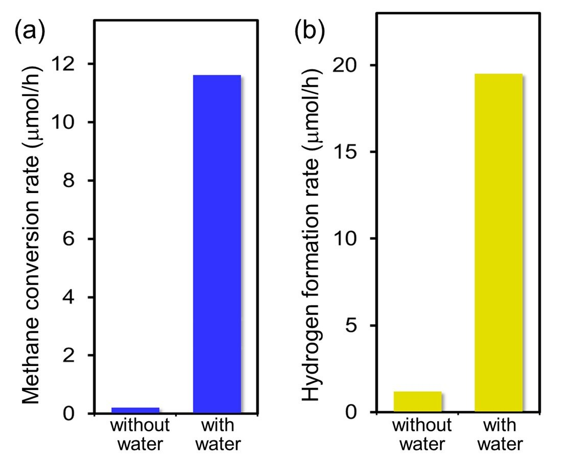
Methane conversion rates and (b) hydrogen formation rates on the Pt/Ga2O3 photocatalysts under ultraviolet irradiation at a methane partial pressure of 70 kPa and water partial pressures of 0 and 2 kPa at a sample temperature of 318 K. The presence of interfacial water significantly enhances the photocatalytic activity at ambient temperatures and pressures. Credit: NINS/IMS

Non-thermal activation and utilization of methane, the main component of natural gas and a ubiquitous natural carbon resource, are among the global challenges for achieving sustainable society. However, incomplete knowledge on microscopic mechanisms of methane activation and hydrogen formation hampers the development of engineering strategies for the reaction system.

Very recently, in a study published in Communications Chemistry, researchers led by Toshiki Sugimoto, Associate Professor at the Institute for Molecular Science, succeeded in obtaining key molecular-level insights into the crucial role of interfacial water on the non-thermal C-H activation in photocatalytic methane conversion. Combining real-time mass spectrometry and operando infrared absorption spectroscopy with ab initio molecular dynamics simulations, they showed that methane conversion is hardly induced by the direct interaction with the trapped hole at the surface Olat site; instead, activation is significantly promoted by low barrier hydrogen abstraction from methane by photoactivated interfacial water species.

In water-mediated processes, the photocatalytic C-H activation is not the rate-determining step, which is in stark contrast to the case of traditional thermocatalytic methane reforming. Moreover, owing to the moderate stabilization of CH3 in the hydrogen-bond network of water, overall photocatalytic conversion rates are dramatically improved by typically more than 30 times at ambient temperatures (~300 K) and pressures (~1 atm). As essentially opposed to thermal catalysis, methane photocatalysis no longer requires high-pressure methane gas (> 20 atm) in the presence of adsorbed water layer.

Critical impacts of interfacial water on C–H activation in photocatalytic methane conversion
Fig. 2. (a) Time evolution of operando infrared (IR) spectra in the O–H stretching region for Pt/Ga2O3 photocatalysts under ultraviolet irradiation at a CH4 pressure of 30 kPa and a D2O pressure of 2 kPa. The O–H peak growth indicates the hydrogen abstraction on catalyst surfaces by photoactivated interfacial water species (CH4(gas) + •OD(ad) → •CH3(ad) + HDO(ad)). (b) Growth rate of the O–H peak (left axis) and CH4 total conversion rate (right axis) on the Pt/Ga2O3 photocatalysts as functions of methane partial pressure; there is a good correlation between the two plots. Credit: NINS/IMS

Water-assisted effects are noticeable also in ethane formation, although water is not explicitly involved in the homocoupling reaction equation (2CH4 → C2H6 + H2). These results indicate that interfacial water kinetically plays crucial roles beyond the traditional thermodynamic concept of redox potential, in which oxidation of water by surface trapped holes is less thermodynamically favored than methane oxidation: E°•OH/H2O = 2.73 V and E°•CH3/CH4 = 2.06 V versus the standard hydrogen electrode.

Notably, these water-assisted effects are commonly observed for several representative photocatalysts with different band-gap energy, such as TiO2, Ga2O3, and NaTaO3, indicating that the incorporation of methane into photoactivated interfacial hydrogen-bond network is essential key for the non-thermal activation of methane.

This work not only expands the molecular-level understanding of the non-thermal C-H activation and conversion, but also provides a fundamental basis for the rational interface design of non-thermal catalytic systems toward the effective and sustainable utilization of methane under ambient conditions.

More information: Critical impacts of interfacial water on C–H activation in photocatalytic methane conversion, Communications Chemistry (2023). DOI: 10.1038/s42004-022-00803-3

Provided by National Institutes of Natural Sciences

Scientists map 3D structure and electronic properties of important biological catalyst

Capturing carbon with both hands
Three-dimensional structure and multiple electron transfer pathways of formate dehydrogenase FoDH1. Credit: Kyoto U/©︎KanonTanaka

Making atoms and electrons behave according to researchers’ intentions is no small task, but scientists often get a little help from nature.

Enzymes from living organisms are well-known for effortlessly directing the buildup and breakdown of molecules in ways that would be difficult or even impossible by conventional chemistry. Putting these biological catalysts to work in industry and health care settings saves time, costs, and even lives.

One such enzyme—FoDH1—is showing great potential for unlocking future carbon capture technologies. Derived from the bacteria Methylorubrum extroquens, this enzyme has the unusual ability to do chemistry with one-carbon molecules, like carbon dioxide. However, its unique electronic properties have left scientists puzzling over its mechanism.

Now a team of Japanese scientists led by Kyoto University has mapped the three-dimensional structure and electronic properties of FoDH1 in unprecedented detail. For the first time, they reveal a unique structure with two sites available for electrons to transfer through metals in the enzyme.

“What’s perhaps even more interesting is that FoDH1 can accept and donate electrons directly, without any other mediating chemicals. This could make it a potential bridge between biological systems and electronic devices,” says corresponding author Keisei Sowa.

In previous work, Sowa’s team hooked the enzyme up to an electrode and demonstrated its unique ability to handle electrons. To further understand how and where electrons were interacting with the enzyme, they examined a single particle of FoDH1 at ultra-low temperatures, revealing the locations of key components, including active clusters of iron and sulfur distributed throughout the enzyme’s structure.

After FoDH1 was probed from different sides with specialized electrodes tipped with gold nanoparticles, signals from two separate sites were unexpectedly detected. Analytical calculations pointed to specific clusters containing different numbers of iron and sulfur atoms that make up the electrode-active sites.

“Our results may be the first clear evidence of two electrode-active sites operating in any enzyme, prompting potential uses of FoDH1 in a range of electrocatalytic processes, including CO2 capture,” reflects Sowa.

“For example, we may be able to see how changing the structure of a mutated enzyme might allow us even greater control of FoDH1’s features.”

The paper, “Multiple electron transfer pathways of tungsten-containing formate dehydrogenase in direct electron transfer-type bioelectrocatalysis,” was published on April 29, 2022 in the journal Chemical Communications.

More information: Tatsushi Yoshikawa et al, Multiple electron transfer pathways of tungsten-containing formate dehydrogenase in direct electron transfer-type bioelectrocatalysis, Chemical Communications (2022). DOI: 10.1039/D2CC01541B

Journal information: Chemical Communications 

Provided by Kyoto University 

In a first, chemists synthesize ocean-based molecule that could fight Parkinson’s

UCLA chemists are first to synthesize ocean-based molecule that could fight Parkinson's
This image illustrates how molecules in nature — in this case, a generic amino acid — can have two enantiomers, or mirror-image versions.  Credit: NASA

Organic chemists at UCLA have created the first synthetic version of a molecule recently discovered in a sea sponge that may have therapeutic benefits for Parkinson’s disease and similar disorders. The molecule, known as lissodendoric acid A, appears to counteract other molecules that can damage DNA, RNA and proteins and even destroy whole cells.

And in an interesting twist, the research team used an unusual, long-neglected compound called a cyclic allene to control a crucial step in the chain of chemical reactions needed to produce a usable version of the molecule in the lab—an advance they say could prove advantageous in developing other complex molecules for pharmaceutical research.

Their findings are published in the journal Science.

“The vast majority of medicines today are made by synthetic organic chemistry, and one of our roles in academia is to establish new chemical reactions that could be used to quickly develop medicines and molecules with intricate chemical structures that benefit the world,” said Neil Garg, UCLA’s Kenneth N. Trueblood Professor of Chemistry and Biochemistry and corresponding author of the study.

A key factor complicating the development of these synthetic organic molecules, Garg said, is called chirality, or “handedness.” Many molecules—including lissodendoric acid A—can exist in two distinct forms that are chemically identical but are 3D mirror images of each other, like a right and left hand. Each version is known as an enantiomer.

When used in pharmaceuticals, one enantiomer of a molecule may have beneficial therapeutic effects while the other may do nothing at all—or even prove dangerous. Unfortunately, creating organic molecules in the laboratory often yields a mixture of both enantiomers, and chemically removing or reversing the unwanted enantiomers adds difficulties, costs and delays to the process.

To address this challenge and quickly and efficiently produce only the enantiomer of lissodendoric acid A that is found almost exclusively in nature, Garg and his team employed cyclic allenes as an intermediate in their 12-step reaction process. First discovered in the 1960s, these highly reactive compounds had never before been used to make molecules of such complexity.

“Cyclic allenes,” Garg said, “have largely been forgotten since their discovery more than half a century ago. This is because they have unique chemical structures and only exist for a fraction of a second when they are generated.”

The team discovered that they could harness the compounds’ unique qualities to generate one particular chiral version of cyclic allenes, which in turn led to chemical reactions that ultimately produced the desired enantiomer of the lissodendoric acid A molecule almost exclusively.

While the ability to synthetically produce an analog of lissodendoric acid A is the first step in testing whether the molecule may possess suitable qualities for future therapeutics, the method for synthesizing the molecule is something that could immediately benefit other scientists involved in pharmaceutical research, the chemists said.

“By challenging conventional thinking, we have now learned how to make cyclic allenes and use them to make complicated molecules like lissodendoric acid A,” Garg said. “We hope others will also be able to use cyclic allenes to make new medicines.”

Co-authors of the research were UCLA doctoral students Francesca Ippoliti (now a postdoctoral scholar at the University of Wisconsin), Laura Wonilowicz and Joyann Donaldson (now of Pfizer Oncology Medicinal Chemistry); UCLA postdoctoral researchers Nathan Adamson and Evan Darzi (now CEO of the startup ElectraTect, a spinoff from Garg’s lab); and Daniel Nasrallah, a UCLA assistant adjunct professor of chemistry and biochemistry.

More information: Francesca M. Ippoliti et al, Total synthesis of lissodendoric acid A via stereospecific trapping of a strained cyclic allene, Science (2023). DOI: 10.1126/science.ade0032

Journal information: Science 

Provided by University of California, Los Angeles 

Cancer-selective chemotherapy using a disassembly-assembly method

A) Chemical structure of Cyto-SA. B) Responsiveness of Cyto-SA towards acidic pH showing no change in the peak position even after 1 h of incubation. CAC determination of Cyto-SA by using Nile red emission method. C)Emission spectra, D) A graph plotting the highest value of each emission spectra. MALDI-TOF/TOF peak of E) 0h incubation and F) 1h incubation. Credit: Advanced Functional Materials (2022). DOI: 10.1002/adfm.202208098

It is well known that cancerous tumor cells have an acidic pH microenvironment (pH 5.6 to 6.8). Using this unique feature, researchers have developed a new anticancer therapeutic agent that selectively kills cancer cells. This access allows detached malignant cells from the tumor to penetrate into cancer cells and induce mitochondrial dysfunction, thereby killing only cancer cells.

This breakthrough has been developed by Professor Ja-Hyoung Ryu and his research team in the Department of Chemistry at UNIST.

In the study, the research team developed transformable nano-assemblies of a mitochondria-targeting agent, Mito-SA, to achieve enhanced tumor selectivity. According to the research team, the new substance formed micelles containing a negative surface charge that selectively disassembled near the tumor cells into parental positively charged Mito-FF molecules, which induced apoptosis via self-assembly into nanofibers in the cancer cell mitochondria.

However, the entry of the Mito-SA micelle inside the normal cell is restricted due to the repulsion between the negatively charged micelle and the negatively charged plasma cell membrane, noted the research team. Furthermore, their findings also revealed that Mito-SA displayed an ideal tumor reduction ability and showed no side effect/damage to the normal tissue.

“Our work shows a strategy for the efficient delivery of positively charged, mitochondria-targeting agents by developing charge-shielded nano-assemblies that selectively disassemble in the tumoral environment,” said the research team.

Their findings have been published in Advanced Functional Materials.

More information: M. T. Jeena et al, Cancer‐Selective Supramolecular Chemotherapy by Disassembly‐Assembly Approach, Advanced Functional Materials (2022). DOI: 10.1002/adfm.202208098

Journal information: Advanced Functional Materials 

Provided by Ulsan National Institute of Science and Technology 

Raman spectroscopy method for rapid identification of beer spoilage bacteria

beer
Credit: Unsplash/CC0 Public Domain

In a study published in Analytical Methods, a research group led by Li Bei from the Changchun Institute of Optics, Fine Mechanics and Physics (CIOMP) of the Chinese Academy of Sciences (CAS) proposed the rapid detection of beer spoilage bacteria based on label-free surface-enhanced Raman spectroscopy (SERS) technology.

Lactic acid bacteria are common spoilage bacteria in beer and need to be monitored and controlled at all stages of beer production. Traditional spoilage bacteria detection methods are time-consuming and cannot meet the demand for real-time, in-situ, rapid detection during the production process.

Raman spectroscopy has been widely used for microbial detection due to its fast, non-destructive and accurate properties, but conventional Raman spectroscopy has the disadvantage of poor signal-to-noise ratio, which affects the accuracy of microbial identification.

Compared with conventional Raman spectroscopy, the SERS technique has a stronger and more sensitive signal and is well suited to the detection of beer spoilage bacteria. Furthermore, the label-free SERS technique is ideal for commercialization due to its low cost and good results.

In this study, the researchers improved the existing process for the preparation of label-free SERS silver nanoparticles (AgNPs). The effect of the AgNPs@KCl agglomeration effect on SERS enhancement was investigated. Eight species of beer spoilage bacteria produced during the beer brewing process were identified by SERS.

The researchers further investigated the effect of the method on the final identification rate by combining the t-distributed stochastic neighbor embedding (t-SNE) dimensionality reduction analysis algorithm, Support Vector Machine (SVM), k-NearestNeighbor (KNN) and Linear Discriminant Analysis (LDA) machine learning algorithms. All three machine learning algorithms achieved an accuracy of around 90% and performed well in identifying beer spoilage bacteria.

In the stability analysis and mixing tests, two known spoilage bacteria were mixed with pure beer and incubated at constant temperature for a period of time to identify the bacteria in the beer. The two spoilage bacteria were successfully detected in the samples and had good spectral stability.

According to the final validation study, the technique can indeed identify the target spoilage bacteria from the simulated samples, which is of great significance to the rapid identification of spoilage bacteria in the beer brewing process.

More information: Lindong Shang et al, Rapid detection of beer spoilage bacteria based on label-free SERS technology, Analytical Methods (2022). DOI: 10.1039/D2AY01221A

Journal information: Analytical Methods 

Provided by Chinese Academy of Sciences 

Researchers develop fluidic memristor with diverse neuromorphic functions

Researchers develop fluidic memristor with diverse neuromorphic functions
Fabrication of PFM. (A) 
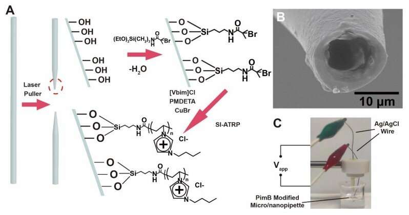
Preparation of PFM with laser puller and SI-ATRP. (B) SEM image of the 10-μm-diameter tip of a PFM. (C) Experimental setup for the electric measurement with the PFM. Credit: Science (2023). DOI: 10.1126/science.adc9150

Neuromorphic devices have attracted increasing attention because of their potential applications in neuromorphic computing, intelligence sensing, brain-machine interfaces and neuroprosthetics. However, most of the neuromorphic functions realized are based on the mimic of electric pulses with solid state devices. Mimicking the functions of chemical synapses, especially neurotransmitter-related functions, is still a challenge in this research area.

In a study published in Science, the research group led by Prof. Yu Ping and Mao Lanqun from the Institute of Chemistry of the Chinese Academy of Sciences developed a polyelectrolyte-confined fluidic memristor (PFM), which could emulate a diverse electric pulse with ultralow energy consumption. Moreover, benefitting from the fluidic nature of PFM, chemical-regulated electric pulses and chemical-electric signal transduction could also be emulated.

The researchers first fabricated the polyelectrolyte-confined fluidic channel by surface-initiated atomic transfer polymerization. By systematically studying the current-voltage relationship, they found that the fabricated fluidic channel could be identified as a memristor, defined as PFM. The origin of the ion memory came from the relatively slow diffusion dynamics of anions into and out of the polyelectrolyte brushes.

The PFM could emulate short-term plasticity patterns (STP), including paired-pulse facilitation and paired-pulse depression. These functions can be operated at a voltage and energy consumption as low as those biological systems, suggesting the potential application in bioinspired sensorimotor implementation, intelligent sensing and neuroprosthetics.

The PFM could also emulate the chemical-regulated STP electric pulses. Based on the interaction between polyelectrolyte and counterions, the retention time could be regulated in different electrolytes. More importantly, in a physiological electrolyte (i.e., phosphate-buffered saline solution, pH 7.4), the PFM could emulate the regulation of memory by adenosine triphosphate (ATP), demonstrating the possibility to regulate the synaptic plasticity by neurotransmitter.

Based on the interaction between polyelectrolytes and counterions, the chemical-electric signal transduction was accomplished with the PFM, which is a key step towards the fabrication of artificial chemical synapses.

With structural emulation to ion channels, PFM features versatility and easily interfaces with biological systems, paving a way to building neuromorphic devices with advanced functions by introducing rich chemical designs. This study provides a new way to interface chemistry with neuromorphic devices.

More information: Tianyi Xiong et al, Neuromorphic functions with a polyelectrolyte-confined fluidic memristor, Science (2023). DOI: 10.1126/science.adc9150

Journal information: Science 

Provided by Chinese Academy of Sciences 

An electrochemical biosensor for antibody detection

An electrochemical biosensor for antibody detection
Graphical Abstract. Credit: Angewandte Chemie International Edition (2022). DOI: 10.1002/anie.202216512

The quantitative detection of specific antibodies in complex samples such as blood can provide information on many different diseases but usually requires a complicated laboratory procedure. A new method for the rapid, inexpensive, yet quantitative and specific point-of-care detection of antibodies has now been introduced in the journal Angewandte Chemie International Edition by an Italian research team. It uses an electrochemical cell-free biosensor that can directly detect antibodies against diseases such as influenza in blood serum.

Influenza is a severe, widespread, epidemic disease that can be fatal and may also have significant societal and economic consequences. The clinical evaluation of immune responses to flu vaccines and infections is thus correspondingly important. A simple, inexpensive, point-of-care diagnostic method would be preferable to current expensive and complex laboratory analysis.

A new method developed by Sara Bracaglia, Simona Ranallo, and Francesco Ricci (University of Rome) fulfills this wish. It is based on “programmable” gene circuits, cell-free transcription, and electrochemical detection.

In living cells, genes are read by RNA polymerases and transcribed into an RNA sequence, which then serves as a blueprint for building proteins (translation). This “machinery” can also be used by cell-free systems. To build their new detector, the team combined this type of machinery with specifically designed synthetic gene circuits that only get “switched on” when the antibody being tested for is present in the sample. As an example, they designed a test that detects anti-influenza antibodies, which are directed against a surface molecule on influenza viruses.

To do this, the team developed a synthetic gene with an incomplete promoter. The promoter is a DNA segment that controls the reading of the gene. If the promoter is incomplete RNA polymerase cannot start the transcription of RNA. The test solution also contains a pair of synthetic DNA strands that are bound to a protein portion (also called peptide) that is specifically recognized by anti-influenza antibodies.

Upon binding between the antibodies and the peptide, the two DNA strands are arranged in a way that completes the promoter and switches the synthetic gene on. The RNA polymerase can now dock on the synthetic gene and start transcribing RNA strands. These RNA strands in turn can bind specifically to a DNA probe fixed to a small disposable electrode and give a measurable current signal change. As long as no antibodies are present, no RNA will be transcribed and no change in current signal will be measured by the disposable electrode. If the sample contains influenza antibodies, the machinery synthesizes RNA, which binds to the electrode leading to a current signal.

The system requires only very small sample volumes, is very specific and sensitive, reliable, and inexpensive. Thus, it can be readily miniaturized to make a portable and easy-to-use diagnostic tool. It is also adaptable for the detection of a wide variety of other antibodies.

More information: Sara Bracaglia et al, Electrochemical Cell‐Free Biosensors for Antibody Detection, Angewandte Chemie International Edition (2022). DOI: 10.1002/anie.202216512

Journal information: Angewandte Chemie International Edition 

Provided by Angewandte Chemie 

Hydrogen storage material’s key restriction identified

Hydrogen storage material's key restriction identified
Just like a “dam burst”, the key bottleneck of MgH2 as hydrogen storage material is the first-layer dehydrogenation. Credit: Royal Society of Chemistry

A group of researchers has identified the key stumbling block of a common solid-state hydrogen material, paving the way for future design guidelines and widespread commercial use.

Details of their findings were published in the Journal of Materials Chemistry A, where the article was featured as a front cover article.

Hydrogen will play a significant role in powering our future. It’s abundant and produces no harmful emissions when burned. But the storage and transportation of hydrogen is both costly and risky.

Currently, hydrogen is stored by three methods: high-pressure gaseous hydrogen storage, low-temperature liquid hydrogen storage, and solid-state hydrogen storage. Among solid-state hydrogen storage, solid-state materials are generally the safest and provide the most hydrogen storage density.

Metal hydrides have long been explored for their large hydrogen storage potentiality and their low cost. As these metals come into contact with gaseous hydrogen, hydrogen gets absorbed onto the surface. Further energy input leads to hydrogen atoms finding their way into the metal’s crystal lattices until the metal becomes saturated with hydrogen. From there, the material can absorb and desorb hydrogen in larger amounts.

Magnesium hydride (MgH2) has shown immense promise for superior hydrogen storage capacity. However, a high temperature is necessary for MgH2 to decompose and produce hydrogen. Furthermore, the material’s complex hydrogen migration and desorption, which result in sluggish dehydrogenation kinetics, have stymied its commercial application.

Hydrogen storage material's key restriction identified
An interesting “burst effect” was found on the dehydrogenation of a typical solid-state hydrogen storage material, MgH2. After the sluggish dehydrogenation at the first layer, hydrogen desorption from the subsequent layers will be much easier. Credit: Hao Li et al.

For decades, scientists have debated why dehydrogenation within MgH2 is so difficult. But now, the research group has uncovered an answer.

Using calculations based on spin-polarized density functional theory with van der Waals corrections, they unearthed a “burst effect” during MgH2‘s dehydrogenation. The initial dehydrogenation barriers measured at 2.52 and 2.53 eV, whereas subsequent reaction barriers were 0.12–1.51 eV.

The group carried out further bond analysis with the crystal orbital Hamilton population method, where they confirmed the magnesium-hydride bond strength decreased as the dehydrogenation process continued.

“Hydrogen migration and hydrogen desorption is much easier following the initial burst effect,” points out Hao Li, associate professor at Tohoku University’s Advanced Institute for Materials Research (WPI-AIMR) and corresponding author of the paper. “Structural engineering tweaks that promote this desorption process could be the key to facilitating the hydrogen desorption of MgH2.”

Li and his colleagues demonstrated that hydrogen vacancies maintained a high degree of electronic localization when the first layer of atomic hydrogen exists. Analyses of the kinetic characteristics of MgH2 after surface dehydrogenation, performed by ab initio molecular dynamics simulations, also provided additional evidence.

“Our findings provide a theoretical basis for the MgH2‘s dehydrogenation kinetics, providing important guidelines for modifying MgH2-based hydrogen storage materials,” adds Li.

More information: Shuai Dong et al, The “burst effect” of hydrogen desorption in MgH2 dehydrogenation, Journal of Materials Chemistry A (2022). DOI: 10.1039/D2TA06458H

Journal information: Journal of Materials Chemistry A 

Provided by Tohoku University 

One-pot reaction creates versatile building block for bioactive molecules

One-pot reaction creates versatile building block for bioactive molecules
Illustration of the new synthetic route for functionalised Z-alkenes. Idea: Dr. Sofiya Runikhina, University of Groningen. Credit: Stutpak Illustration and Animation, Deventer, the Netherlands

Chemists from the University of Groningen have found a simple way to produce previously inaccessible chiral Z-alkenes, molecules that offer a significant synthetic short-cut for the production of bioactive molecules.

Instead of eight to ten synthetic steps to produce these molecules, the new reaction can be done in three steps, without the need for any purification. The key lies in a phosphine molecule that is normally used to make metal-containing catalysts but that turns out to be the ideal starting point for this chemical reaction. The results were published in Science Advances on January 13.

Organic compounds are very versatile. Their carbon atoms can be connected by single, double, or triple bonds. Furthermore, many biologically important molecules contain chiral centers, parts of the molecule that can be in two mirror image positions, comparable to a left and a right hand. Molecules that have a double bond, a chiral center, and a reactive group for synthetic modifications all next to each other are also important, but chemists find that these are very difficult to make.

Unstable

Alkenes are compounds that contain two carbon atoms that are connected by a double bond. When picturing these two carbon atoms horizontally, we can distinguish Z-alkenes, whereby both carbon atoms are connected to another carbon on the same side (both pointing up), and E-alkenes, whereby the connected carbons are on opposite sides, (one up and one down). Z-alkenes are unstable because carbons that are connected on the same side are forced to be close to each other.

“The molecule doesn’t like this and if it gets a chance it will transform into the more stable E-alkene. That is why it is hard to make less stable Z-configured alkenes,” explains Syuzanna Harutyunyan, Professor of Homogeneous Catalysis at the University of Groningen. “Z-alkenes are very useful, but also difficult to make.”

The team needed to make the less stable Z-alkenes, where the double bond is connected to a chiral carbon center, further connect to a highly reactive carbon center, which is very tricky.

One-pot reaction creates versatile building block for bioactive molecules
This picture shows the configuration of Z- and E-alkenes (upper panel) and the one-pot synthesis of functionalised Z-alkenes. Credit: Harutyunyan Lab, University of Groningen

Reactive salt

Using known synthetic methods, it would take about eight to ten separate steps to create such a structure. Harutyunyan and her team tried to simplify this by starting out with a molecule called phosphine. Co-author Roxana Postolache says, “This molecule is normally used to produce metal-containing catalysts. In previous work, we developed a way to make a chiral phosphine, which formed the basis for our new synthetic route to Z-alkenes.”

Harutyunyan states, “We took our phosphine and turned it into a salt. This would allow the creation of a double bond with Z-configuration.”

But this salt is very reactive and all the attempts to introduce a double bond resulted in a lot of products that the scientists did not want. “So, we had to find a way to tune the reactivity,” explains Postolache.

Blackboard

This step required a blackboard and chalk approach, which Harutyunyan and her team used to discuss options. A potential solution was found in adding a special group to the phosphine to make a different type of salt. Harutyunyan says, “We figured that this should pull electrons away from the phosphorus and would allow us to tune the reactivity.”

First author Luo Ge took the idea from the blackboard to the laboratory. “We tried to make this idea work and we got it right with our first attempt. It was a pleasant surprise to see that our idea really worked.” They subsequently optimized the reaction and then used their method to modify real bioactive compounds.

Possibilities

A big advantage of the new synthetic route is that it takes fewer steps and is essentially a one-pot reaction. It just requires room temperature for the first step, mild heating (50–70 °C) to make the salt, and –78 °C for the final step of making the double bond with a Z-configuration.

Joint first author Esther Sinnema says, “By using our phosphine as a synthetic tool, rather than a catalyst, we opened up all kinds of possibilities. We could make a large number of new chiral Z-alkenes and use the method to modify bioactive compounds. In the paper, we present 35 different molecules that were made with our method.”

“We expect that our study will pave the way for using commercially available, simple alkenes to make much more complex functionalized alkenes via phosphine and salt intermediates,” says Harutyunyan.

More information: Luo Ge et al, Enantio- and Z-selective synthesis of functionalized alkenes bearing tertiary allylic stereogenic center, Science Advances (2023). DOI: 10.1126/sciadv.adf8742www.science.org/doi/10.1126/sciadv.adf8742

Journal information: Science Advances 

Provided by University of Groningen