Chemists develop new method to create chiral structures

by University of California – Riverside

Chemists develop new method to create chiral structures
The photos depict the vibrant colors exhibited by a dispersion of magnetic nanoparticles when subjected to magnetic fields with varying chiral distributions, as observed through polarized lenses. Credit: Yin lab, UC Riverside.

Some molecules exist in two forms, such that their structures and their mirror images are not superimposable, like our left and right hands. Called chirality, it is a property these molecules have due to their asymmetry. Chiral molecules tend to be optically active because of how they interact with light. Oftentimes, only one form of a chiral molecule exists in nature, for example, DNA. Interestingly, if a chiral molecule works well as a drug, its mirror image could be ineffective for therapy.

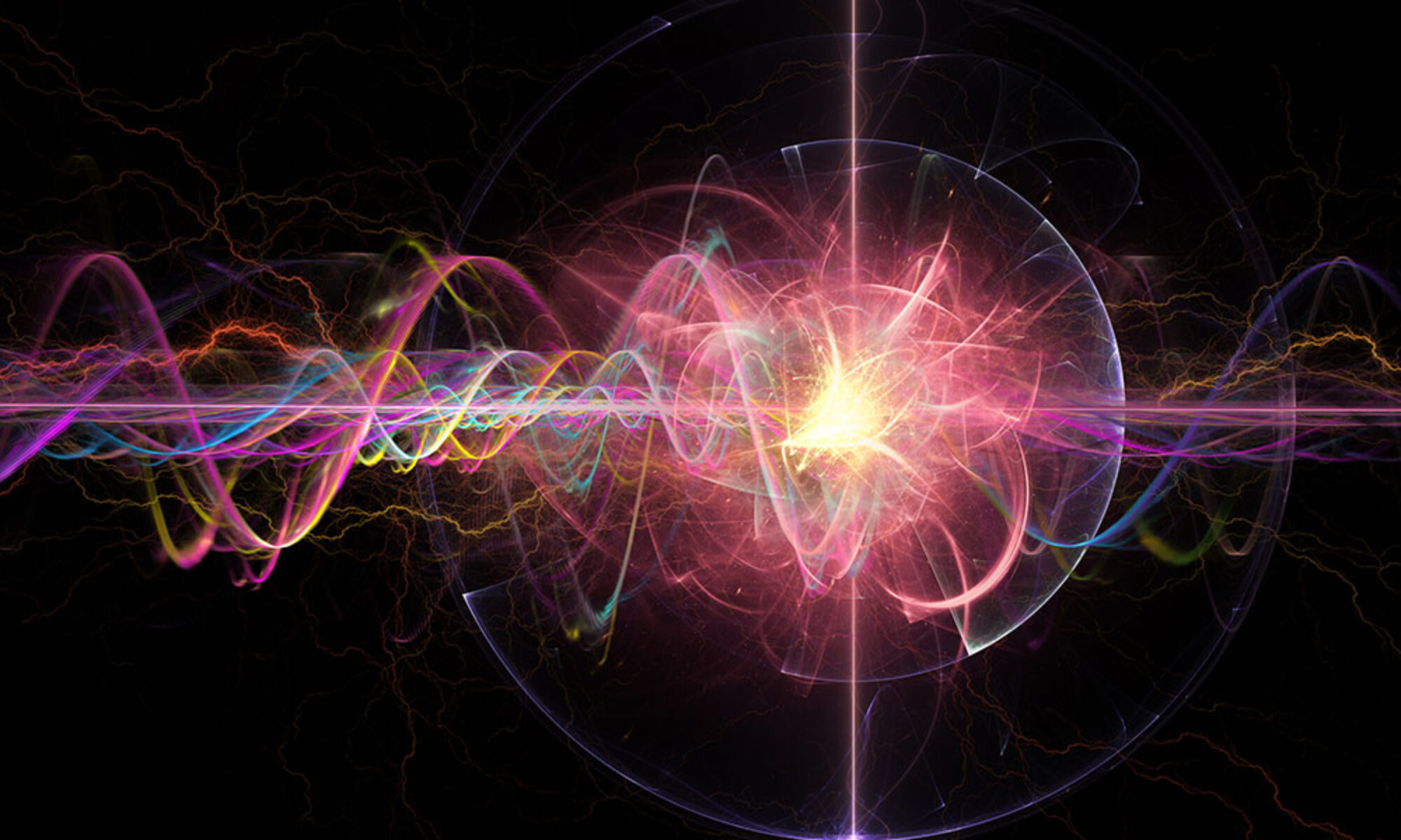
In trying to produce artificial chirality in the lab, a team led by chemists at the University of California, Riverside, has found that the distribution of a magnetic field is itself chiral. The research paper titled, “A magnetic assembly approach to chiral superstructures,” appears in the journal Science.

“We discovered that the magnetic field lines produced by any magnet, including a bar magnet, have chirality,” said Yadong Yin, a professor of chemistry, who led the team. “Further, we were also able to use the chiral distribution of the magnetic field to coax nanoparticles into forming chiral structures.”

Traditionally, researchers have used “templating” to create a chiral molecule. A chiral molecule is first used as the template. Achiral (or non-chiral) nanoparticles are then assembled on this template, allowing them to mimic the structure of the chiral molecule. The drawback to this technique is that it cannot be universally applied, being heavily dependent on the specific composition of the template molecule. Another shortcoming is the newly formed chiral structure cannot be easily positioned at a specific location on, say, an electronic device.

“But to gain an optical effect, you need a chiral molecule to occupy a particular place on the device,” Yin said. “Our technique overcomes these drawbacks. We are able to rapidly form chiral structures by magnetically assembling materials of any chemical composition at scales ranging from molecules to nano- and microstructures.”

Yin explained that his team’s method uses permanent magnets that consistently rotate in space to generate the chirality. He said transferring chirality to achiral molecules is done by doping, that is incorporating guest species, such as metals, polymers, semiconductors, and dyes into the magnetic nanoparticles used to induce chirality.

Yin said chiral materials acquire an optical effect when they interact with polarized light. In polarized light, light waves vibrate in a single plane, reducing the overall intensity of the light. As a result, polarized lenses in sunglasses cut glare to our eyes, while non-polarized lenses do not.

“If we change the magnetic field that produces a material’s chiral structure, we can change the chirality, which then creates different colors that can be observed through the polarized lenses,” Yin said. “This color change is instantaneous. Chirality can also be made to disappear instantaneously with our method, allowing for rapid chirality tuning.”

The findings could have applications in anti-counterfeit technology. A chiral pattern that signifies the authenticity of an object or document would be invisible to the naked eye but visible when seen through polarized lenses. Other applications of the findings are in sensing and the field of optoelectronics.

“More sophisticated optoelectronic devices can be made by taking advantage of the tunability of chirality that our method allows,” said Zhiwei Li, the first author of the paper and former graduate student in Yin’s lab. “Where sensing is concerned, our method can be used to rapidly detect chiral or achiral molecules linked to certain diseases, such as cancer and viral infections.”

Yin and Li were joined in the research by a team of graduate students in Yin’s lab, including Qingsong Fan, Zuyang Ye, Chaolumen Wu, and Zhongxiang Wang. Li is now a postdoctoral researcher at Northwestern University in Illinois.

More information: Zhiwei Li et al, A magnetic assembly approach to chiral superstructures, Science (2023). DOI: 10.1126/science.adg2657www.science.org/doi/10.1126/science.adg2657

Journal information: Science 

Provided by University of California – Riverside 

Researchers create edible, transparent composite packaging with biocellulose

by Society of Chemical Industry

Researchers create edible, transparent composite packaging with biocellulose
By incorporating soy protein into the structure and coating it with an oil-resistant composite, the CUHK team successfully created an edible, transparent, and robust BC-based composite packaging. Credit: To Ngai

Plastic food packaging accounts for a significant proportion of plastic waste in landfills. In the face of escalating environmental concerns, researchers are looking to bio-derived alternatives.

Now, scientists at The Chinese University of Hong Kong (CUHK) have developed an edible, transparent and biodegradable material with considerable potential for application in food packaging. Their work is published in the Journal of the Science of Food and Agriculture.

Heavy reliance on petrochemicals and inherent non-biodegradability of plastic packaging mean it has long been a significant contributor to environmental contamination. A team at CUHK has turned its attention to bacterial cellulose (BC), an organic compound derived from certain types of bacteria, which has garnered attention as a sustainable, easily available, and non-toxic solution to the pervasive use of plastics.

Professor To Ngai from the Department of Chemistry, CUHK and corresponding author of the study, explained that the impressive tensile strength and high versatility of BC are the key to its potential.

He said, “Extensive research has been conducted on BC, including its use in intelligent packaging, smart films, and functionalized materials created through blending, coating, and other techniques. These studies demonstrate the potential of BC as a replacement for single-use plastic packaging materials, making it a logical starting point for our research.”

Unlike the cellulose found in the cell walls of plants, BC can be produced through microbial fermentation, which eliminates the need for harvesting trees or crops. Ngai noted that as a result, “…this production method does not contribute to deforestation or habitat loss, making BC a more sustainable and environmentally friendly material alternative to plant cellulose.”

Up until now, the widespread adoption of BC has been limited by its unfavorable sensitivity to moisture in the air (hygroscopicity), which detrimentally impacts its physical properties.

In the paper, the researchers laid out a novel approach to address the limitations of BC-based materials. By incorporating certain soy proteins into the structure and coating it with an oil-resistant composite, they successfully created an edible, transparent, and robust BC-based composite packaging.

Researchers create edible, transparent composite packaging with biocellulose
Bacterial cellulose (BC) – an organic compound derived from certain types of bacteria which has garnered attention as a sustainable, easily available, and non-toxic solution to the pervasive use of plastics. Credit: To Ngai

Ngai noted that this approach has a high feasibility for scale-up: “It does not require specific reaction conditions like chemical reactions, but rather a simple and practical method with mixing and coating. This approach offers a promising solution to the challenge of developing sustainable and environmentally friendly packaging materials that can replace single-use plastics on a large scale.”

The study demonstrated that the plastic alternative could be completely degraded within 1-2 months. Unlike other bio-derived plastics such as polylactic acid, the BC-based composite does not require specific industrial composting conditions to degrade.

Ngai explained, “The material developed in this research is completely edible, making it safe for turtles and other sea animals to consume without causing aquatic toxicity in the ocean.”

The researchers at CUHK are now exploring the directions for future research. They hope to enhance the versatility of modified BC films, making them suitable for a wider range of applications. Specifically, they are focused on developing a thermosetting glue that can create strong bonds between bacterial cellulose, allowing it to be easily molded into various shapes when heated.

“One of the main challenges with bacterial cellulose films is that they are not thermoplastic, which limits their potential for use in certain applications. By addressing this issue, we hope to make bacterial cellulose films more competitive with traditional plastics while maintaining their eco-friendliness,” explained Ngai.

Ngai hopes that the current study will help to combat the excessive use of single-use plastics, which can persist for hundreds of years after only a few days of being displayed on supermarket shelves.

“This research serves as a reminder that natural raw materials may already possess the necessary characteristics to perform beyond the functions of plastic packaging,” he concluded.

More information: Ka Man Cheung et al, Edible, strong, and low‐hygroscopic bacterial cellulose derived from biosynthesis and physical modification for food packaging, Journal of the Science of Food and Agriculture (2023). DOI: 10.1002/jsfa.12758

Journal information: Journal of the Science of Food and Agriculture 

Provided by Society of Chemical Industry

Unraveling the role of the NiO electrocatalyst in alcohol electrooxidation reactions

Unraveling the catalytic role of NiO electrocatalyst in alcohol electrooxidation reaction
The synergy between EOM-HAT and the hydration of aldehyde results in the electrooxidation of R-CH2OH to R-COOH. The synergy between EOM-HAT and EOM-Cleavage of the C-C bond causes the electrooxidation of R-CHOH-CH2OH to R-COOH and HCOOH. Credit: Science China Press

A study led by Dr. Wei Chen, Prof. Yuqin Zou, and Prof. Shuangyin Wang (State Key Laboratory of Chemo/Bio-Sensing and Chemometrics, College of Chemistry and Chemical Engineering, Advanced Catalytic Engineering Research Center of the Ministry of Education, Hunan University) unravels the reaction mechanism of the primary alcohol/vicinal diol electrooxidation reaction on NiO, especially for the synergy between the electrochemical and non-electrochemical steps.

The alcohol electrooxidation on NiO is an indirect electrooxidation reaction with the electrophilic oxygen species as the redox mediator. Therefore, the electrochemical step is the electrochemical generation of electrophilic oxygen species. The research team found two kinds of NiO electrocatalyst function mechanisms, i.e., the electrophilic oxygen-mediated mechanism involving hydrogen atom transfer (EOM-HAT) and the electrophilic oxygen-mediated mechanism involving C-C bond cleavage (EOM-Cleavage of C-C bond).

The synergy between EOM-HAT and the hydration of aldehyde results in the electrooxidation of primary alcohol (R-CH2OH) to carboxylic acid (R-COOH) on NiO. On the other hand, the synergy between EOM-HAT and EOM-Cleavage of the C-C bond causes the electrooxidation of vicinal diol (R-CHOH-CH2OH) to R-COOH and formic acid (HCOOH) on NiO.

This study, published in the journal National Science Review, highlights the synergy between the electrochemical and non-electrochemical steps in the alcohol electrooxidation reaction. It establishes a unified reaction mechanism of alcohol electrooxidation reaction based on nickel-based electrocatalysts.

More information: Wei Chen et al, Unraveling the electrophilic oxygen-mediated mechanism for alcohol electrooxidation on NiO, National Science Review (2023). DOI: 10.1093/nsr/nwad099

Provided by Science China Press 

Multifunctional self-healing liquid metal hydrogel developed for human-computer interaction

Multifunctional self-healing liquid metal hydrogel developed for human-computer interaction
Schematic structure and application of the liquid metal hydrogel. Credit: Li XIaofei

Recently, researchers from the Hefei Institutes of Physical Science (HFIPS) of the Chinese Academy of Sciences, led by Prof. Tian Xingyou and Prof. Zhang Xian, along with associate Prof. Yang Yanyu from the College of Materials Science and Engineering at Zhengzhou University, used gallium indium alloy (EGaIn) to initiate the polymerization and serve as flexible fillers to construct liquid metal/polyvinyl alcohol(PVA)/P(AAm-co-SMA) double network hydrogel.

“The resulting material was super-stretchable and self-healing,” said Li Xiaofei, first author of the paper, “it will promote the research and practical application of hydrogels and liquid metal in intelligent devices and military fields.”

The study was published in Materials Horizons.

Most conductive hydrogels suffer from subpar mechanical qualities and lack desirable self-recovery and self-healing abilities, severely limiting hydrogels’ potential uses. Liquid metals like gallium indium alloy (EGaIn) can toughen polymers by conforming to their changing shapes. Also, gallium (Ga) in EGaIn can initiate vinyl monomer’s free radical polymerization.

In this research, the team built a liquid metal/PVA/P(AAm-co-SMA) double network hydrogel (LM hydrogel) with EGaIn serving as both the polymerization initiator and the flexible fillers.

The PVA network used PVA microcrystals and coordination interaction of Ga3+ and PVA as cross-links, while the P(AAm-co-SMA) network used hydrophobic association and the EGaIn microspheres. The LM hydrogel was endowed with excellent super-stretchability (2000%), toughness (3.00 MJ/m3), notch resistance, and self-healing property (> 99% at 25 °C after 24 h) due to the multiple physical cross-links and the synergistic effect of the rigid PVA microcrystal network and the ductile P(AAm-co-SMA) hydrophobic network.

“The sensors developed for it can be used in health monitoring and motion identification through human-computer interaction,” said Li Xiaofei, “thanks to the LM hydrogel’s sensitive strain sensing capability.”

As a result of EGaIn’s low infrared emissivity and remarkable photothermal, LM hydrogel shows considerable promise in infrared camouflage.

More information: Xiaofei Li et al, Self-healing liquid metal hydrogel for human–computer interaction and infrared camouflage, Materials Horizons (2023). DOI: 10.1039/D3MH00341H

Journal information: Materials Horizons 

Provided by Chinese Academy of Sciences 

Researchers discover a new way to develop drugs without side effects

A new way to develop drugs without side effects
An artist’s impression of the GPCR activation from inside the cell and the resulting targeted response. Credit: Kobayashi and Kawakami et al., 2023

Have you ever wondered how drugs reach their targets and achieve their function within our bodies? If a drug molecule or a ligand is a message, an inbox is typically a receptor in the cell membrane. One such receptor involved in relaying molecular signals is a G protein-coupled receptor (GPCR). About one-third of existing drugs work by controlling the activation of this protein. Japanese researchers now reveal a new way of activating GPCR by triggering shape changes in the intracellular region of the receptor. This new process can help researchers design drugs with fewer or no side effects.

If the cell membrane is like an Oreo cookie sandwich, GPCR is like a snake with seven segments traversing in and out of the cookie sandwich surface. The extracellular loops are the inbox for messages. When a message molecule binds to the extracellular side of the receptor, it triggers a shape change activating G proteins and the ß-arrestin protein attached to the intracellular side of the receptor. Like a molecular relay, the information passes downstream and affects various bodily processes. That is how we see, smell, and taste, which are sensations of light, smell, and taste messages.

Adverse side effects ensue if drugs acting on GPCRs activate multiple signaling pathways rather than a specific target pathway. That is why drug development focuses on activating specific molecular signal pathways within cells. Activating the GPCR from inside the cell rather than outside the cell could be one way to achieve specificity. But until now, there was no evidence of direct activation of only the intracellular side of GPCRs without the initiations from the extracellular side.

A team of researchers headed by Osamu Nureki, a professor at the University of Tokyo, and his lab, discovered a new receptor activation mode of a bone metabolism-related GPCR called human parathyroid hormone type 1 receptor (PTH1R) without signal transduction from the extracellular side.

“Understanding the molecular mechanism will enable us to design optimal drugs,” says Kazuhiro Kobayashi, a doctoral student and an author of the study. Such a drug offers “a promising treatment for osteoporosis.”

Kobayashi has been conducting research on bone formation in animal models since he was an undergrad. “Treatments for osteoporosis that target PTH1R require strict dosage, have administrative restrictions, and there aren’t yet any better alternatives,” he says. That motivated their team to look for better drug design strategies targeting the parathyroid hormone receptor.

To understand function through structure, they used cryo-electron microscopy and revealed the 3D structure of the PTH1R and G protein bound to a message molecule. The team synthesized a non-peptide message molecule called PCO371 which binds to the intracellular region of the receptor and interacts directly with G protein subunits. In other words, PCO371 activates the receptor after entering the cell.

The PCO371-bound PTH1R structure can directly and stably modulate the intracellular side of PTH1R. And because PCO371 activates only G protein and not ß-arrestin it does not cause side effects. This specificity of its binding and receptor activation mode makes it a suitable candidate for potential small-molecule-based drugs for class B1 GPCRs, like PTH1R, which currently lack oral administrative drug ligands. Such drugs would have reduced adverse effects and burdens on patients as they act on specific molecular pathways.

The findings from this study will help “develop new drugs for disorders such as obesity, pain, osteoporosis, and neurological disorders.”

The study appears in the journal Nature.

More information: Kazuhiro Kobayashi et al, Class B1 GPCR activation by an intracellular agonist, Nature (2023). DOI: 10.1038/s41586-023-06169-3

Journal information: Nature 

Provided by University of Tokyo 

Functional analyses of RNA-related enzymes using a next-generation DNA sequencer

tRNA-MaP: functional analyses of RNA-related enzymes using a next-generation DNA sequencer
Overview of tRNA-MaP. Credit: Ehime University, Ryota Yamagami, Hiroyuki Hori

Genetic information encoded in genomic DNA is transcribed to mRNAs and then the codons on mRNA are decoded by transfer RNAs (tRNAs) during protein synthesis. tRNAs deliver amino acids to ribosomes and proteins are synthesized from the amino acids on the ribosomes according to the decoded genetic information. Therefore, tRNA plays a key role during the translation of genetic information.

tRNAs contain numerous modified nucleosides, which regulate the accuracy and efficiency of protein synthesis. Modified nucleosides in tRNA are synthesized by tRNA modification enzymes. Therefore, unveiling the mechanisms by which tRNA modification enzymes selectively recognize substrate tRNAs from non-substrate RNAs; the when, where, and how many tRNAs are being modified by the modification enzymes, is of crucial importance to understand the protein synthesis machinery.

Addressing these key questions is, however, challenging due to the lack of a high-throughput technique that identifies the characteristic properties of tRNA modification enzymes.

To overcome this issue, Drs. Yamagami and Hori at Ehime University applied next-generation DNA sequencing technology to functional analyses of tRNA modification enzymes and developed a new high-throughput assay method, “tRNA-MaP.”

The tRNA-MaP technique can rapidly screen an RNA pool consisting of more than 5,000 RNA species and identify the substrate tRNAs of the target tRNA modification enzyme(s) with comparative sensitivity to already-established methods. By tRNA-MaP, in combination with protein orthology analyses, the researchers predicted numerous natural modifications in Geobacillus stearothermophilus tRNAs.

Furthermore, they analyzed the substrate recognition mechanism of G. stearothermophilus tRNA m1A22 methyltransferase (TrmK), which methylates adenosine at position 22 to 1-methyladenosine (m1A22) in tRNA, using tRNA-Map. Mutation profiling has revealed that TrmK selects a subset of tRNAs for the substrate.

Using 240 variants of G. stearothermophilus tRNALeu transcripts, the researchers found that U8, A14, G15, G18, G19, U55, Purine57 and A58 are important for the methylation by TrmK. In addition, based on the recognition sites in tRNA and the crystal structure of TrmK, a docking model between TrmK and tRNA has been constructed.

This study, now published in the Journal of Biological Chemistry, revealed that tRNA-Map is applicable for the analysis of the tRNA modification enzyme. Notably, because tRNA-Map can analyze any RNA molecular species from any organism, even DNA molecules, tRNA-Map can be used for analysis of all nucleic acid-related proteins except for tRNA modification enzymes. Thus, tRNA-Map can accelerate the integrative understanding of the flow of genetic information.

More information: Ryota Yamagami et al, Application of mutational profiling: New functional analyses reveal the tRNA recognition mechanism of tRNA m1A22 methyltransferase, Journal of Biological Chemistry (2022). DOI: 10.1016/j.jbc.2022.102759

Journal information: Journal of Biological Chemistry 

Provided by Ehime University

Building better catalysts to close the carbon dioxide loop

Building better catalysts to close the carbon dioxide loop
Graphical abstract. Credit: ACS Catalysis (2022). DOI: 10.1021/acscatal.2c03055

The best way to stave off the worst effects of climate change is to reduce CO2 emissions around the world. And one way to do that, says Zhongwei Chen, a professor in the Department of Chemical Engineering at the University of Waterloo, is to capture the CO2 and convert it into other useful chemicals, such as methanol and methane for fuels.

Stopping emissions at the source, and further reducing future ones by replacing CO2-producing fuels with cleaner ones “…is a way to close the circle,” Chen says.

In order to turn CO2 into methanol, you need a catalyst to jump-start the electrochemical reaction. Traditionally, these catalysts have either been made out of precious metals like gold or palladium, or base metals like copper or tin. However, they are expensive and break down easily, hindering large-scale implementation.

“Right now we can’t meet industrial requirements,” says Chen. “So we are trying to design catalysts with better activity, selectivity, and durability.”

Chen and his team are focused on low-cost metal and metal-free catalysts. The metal-free catalysts, made from carbon, are cheaper and more durable but tend to have lower catalytic activity than metal ones. So the team tweaked the chemical composition and physical design of the catalyst to optimize its efficiency, combining the materials science of the catalyst design with the engineering of the electrode and reactor to improve the activity of the whole system.

“We want to make it as small as we can, but not too small to be a practical application,” says Chen. They combined nanometer-scale active sites within a micrometer-scale particle—like bubbles in a tiny sponge—to create a catalyst with a huge number of active sites in a particle that is easy and practical to fabricate.

The powerful light beams and expert technical teams at the Canadian Light Source (CLS) at the University of Saskatchewan were instrumental in helping to design an efficient catalyst, says Chen. “The advanced facilities at CLS are critical in helping us understand what was going on during the reaction, so we can continue to design and improve the next generation of catalysts.”

The paper is published in the journal ACS Catalysis.

More information: Zhen Zhang et al, Steering Carbon Hybridization State in Carbon-Based Metal-free Catalysts for Selective and Durable CO2 Electroreduction, ACS Catalysis (2022). DOI: 10.1021/acscatal.2c03055

Journal information: ACS Catalysis 

Provided by Canadian Light Source 

Could a new test that detects dopamine levels help diagnose neurological diseases?

dopamine
Ball-and-stick model of the dopamine molecule, a neurotransmitter that affects the brain’s reward and pleasure centers. Credit: Jynto/Wikipedia

Altered levels of the neurotransmitter dopamine are apparent in various conditions, such as Parkinson’s disease and depression.

In research published in ChemistrySelect, investigators describe a quick, sensitive, and simple test to determine dopamine levels in biological fluids. The method could help clinicians spot abnormal blood levels of dopamine in patients, potentially allowing for earlier disease detection.

The method relies on what are called carbon quantum dots, a type of carbon nanomaterial with photoluminescence properties, and ionic liquid, which is comprised of several mineral anions and organic cations existing in liquid form at room temperature.

“The proposed electrochemical sensor could be an exceptional step forward in dopamine detection and pave the way for the molecular diagnosis of neurological illnesses,” the authors wrote.

Could a new test that detects dopamine levels help diagnose neurological diseases?
Credit: ChemistrySelect (2023). DOI: 10.1002/slct.202203630

More information: Zahra Nazari et al, An Electrochemical Sensor Based on Carbon Quantum Dots and Ionic Liquids for Selective Detection of Dopamine, ChemistrySelect (2023). DOI: 10.1002/slct.202203630

Provided by Wiley 

Wireless sensor enables real-time spoilage alerts on food

Revolutionary sensor enables real-time spoilage alerts on food
Summary of the concept. Credit: Nature Food (2023). DOI: 10.1038/s43016-023-00750-9

Food waste and food-borne diseases are among the most critical problems urban populations face today. They contribute to greenhouse emissions tremendously and amplify economic and environmental costs. Since food spoilage remains the main reason for this waste, the circumstances of processing, transporting, and preserving food still need to be improved in line with current technological advancements.

Current monitoring processes are conducted in laboratories and use expensive chromatographic devices. These not only require too much time but also excessive resources and qualified personnel. So, present methods unfortunately prove to be inefficient in today’s circumstances.

New research published in Nature Food presents a significant alternative to this process: A new user-friendly, cost-effective, and up-to-date sensor that can be applied on food directly and replace lab-monitoring. The 2 x 2 cm miniature wireless device introduced in the paper offers real-time measurement, is battery-free and smartphone-compatible. It is expected to be highly effective especially in high-protein foods such as beef, chicken, and fish.

The research was led by Dr. Emin İstif (Molecular Biology and Genetics, Kadir Has University) and Asst. Prof. Levent Beker (Mechanical Engineering, Koç University) with the contribution of Prof. İskender Yılgör and Dr. Emel Yılgör (Chemistry, Koç University), Asst. Prof. Çağdaş Dağ (Molecular Biology and Genetics, Koç University) and Asst Prof. Hatice Ceylan Koydemir (Texas A&M University).

While existing solutions focus on the change in color of food, this new device, for the first time, offers a capacitive measurement method and thus utilizes near-field communication (NFC) technology with power-free and wireless communication. The authors indicate that this eliminates major disadvantages encountered in resistive devices such as moisture sensitivity and incorrect data due to distance.

The invention will not only provide companies the opportunity of reducing costs but also help consumers tremendously. Once widely commercialized, the device will enable continuous monitoring on shelves and allow users to control freshness right before buying a product or even before consumption at home. This opportunity of on-demand spoilage analysis via mobile phones will ultimately help preventing food waste and food-borne diseases.

With its cost-effectiveness and accessibility, the authors hope to contribute to the greater struggle against global warming and greenhouse emissions more effectively and quickly. The next steps will be to focus on increasing the potential for commercialization of the product in the near future.

More information: Emin Istif et al, Miniaturized wireless sensor enables real-time monitoring of food spoilage, Nature Food (2023). DOI: 10.1038/s43016-023-00750-9

Journal information: Nature Food 

Provided by Koc University 

A poison helps us understand molecular hydrogen-producing biocatalysts

A poison helps to understand molecular hydrogen-producing biocatalysts
Superimposition of structures of CpI in various states. Credit: Angewandte Chemie International Edition (2022). DOI: 10.1002/anie.202216903

In nature, enzymes termed hydrogenases are capable of producing molecular hydrogen (H2). Special types of these biocatalysts, so-called [FeFe]-hydrogenases, are extremely efficient and therefore of interest for biobased hydrogen production. Although scientists have learned a lot about how these enzymes work, many details remain to be completely understood.

A research team of the Photobiotechnology group at Ruhr University Bochum, Germany, headed by Dr. Jifu Duan and Professor Thomas Happe succeeded in filling a scientific gap. The researchers showed that external cyanide binds to the [FeFe] hydrogenases and inhibits hydrogen formation. In the process, they detected a structural change in the proton transport pathway, which helps to understand the coupling of electron and proton transport. They reported their findings in the journal Angewandte Chemie of December 4, 2022.

A sophisticated internal catalyst

To generate H2, these biocatalysts transfer electrons to protons, employing a sophisticated structure as internal catalyst. This so-called H-cluster contains electronically active iron ions that are bound to what most people know as toxins: carbon monoxide and cyanide.

However, although internal carbon monoxide and cyanide are crucial for the high activity of hydrogenases, additional external carbon monoxide binds to the H-cluster and prevents its H2 production. “Interestingly, cyanide is also a well-known inhibitor of iron-containing biocatalysts,” says Jifu Duan. “And yet, its effect on [FeFe]-hydrogenases has hardly been analyzed before.”

The Bochum-based research team closed this scientific gap. The researchers showed that external cyanide binds to and inhibits [FeFe]-hydrogenases. In collaboration with Professor Eckhard Hofmann, head of the protein crystallography group at RUB, the team obtained the structure of H2-producing biocatalysts to which external cyanide was bound.

“The high-resolution structure in combination with spectroscopic analyses tells us that the external cyanide directly binds to the H-cluster, similar to other inhibitors studied so far,” says Jifu Duan. “This explains why the hydrogenase is inactive after cyanide treatment.”

Coincidental capture of a transient state

When the researchers took a detailed look into the structure of the cyanide-poisoned hydrogenase, they found a surprise. They observed structural changes in the proton transport pathway that is required to guide the protons that will become H2 to the H-cluster.

“This conformation has been suggested to be vital for efficient proton shuttling, but it had never been observed structurally. Coincidently, the cyanide binding helped us to capture such a transient state,” says Jifu Duan.

“These findings are important for researchers to understand the coupling of electron and proton transport which is not only relevant for H2-generating enzymes, but many additional biocatalysts,” concludes Thomas Happe.

More information: Jifu Duan et al, Cyanide Binding to [FeFe]‐Hydrogenase Stabilizes the Alternative Configuration of the Proton Transfer Pathway, Angewandte Chemie International Edition (2022). DOI: 10.1002/anie.202216903

Journal information: Angewandte Chemie International Edition  Angewandte Chemie 

Provided by Ruhr-Universitaet-Bochum