Novel computational approach reveals previously inaccessible druggable pockets

by University of Amsterdam

Decrypting integrins by mixed-solvent molecular dynamics simulation
Credit: HIMS

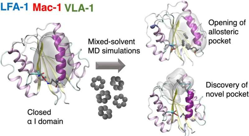
A team of researchers from the Universities of Amsterdam and Zurich together with the Swiss company Allocyte Pharmaceuticals have for the first time been able to discover allosteric sites in a type of cell surface receptor called integrin.

In a paper recently published in the Journal of Chemical Information and Modeling, they describe how this reveals previously inaccessible druggable integrin pockets. At the heart of the research is a novel computational approach for mixed-solvent molecular dynamics simulation developed by Dr. Ioana Ilie at the Computational Chemistry group of the Van ‘t Hoff Institute for Molecular Sciences at the University of Amsterdam.

Integrins are a family of cell surface adhesion receptors which are capable of transmitting signals bidirectionally across membranes. They are known for their therapeutic potential in a wide range of diseases. However, the development of integrin targeting medication has been impacted by unexpected downstream effects. In particular, these are observed with drugs targeting the native binding site of the integrin.

The so-called allosteric modulation of integrins is a promising approach to potentially overcome these limitations. Here, the drug binds elsewhere on the receptor, changing the conformation and thus impacting the activity of the protein. Allosteric modulation of receptors therefore creates opportunities for drug discovery and development which are potentially superior to classic orthosteric modulation.

Novel druggable pockets

The novel computational approach developed by Ioana Ilie relies on enriching the solvent with small organic molecules (benzene in small concentrations) to enable the gentle opening of the integrin α I domain. This revealed novel previously inaccessible druggable pockets within the integrins LFA-1, VLA-1, and Mac-1. This study thus offers structural and dynamic insight on the effect of small alterations in solvent conditions on the accessibility of novel potentially druggable pockets, which are validated via virtual screening. The study acts as proof of concept and sets the foundation for the design of the next-generation integrin-targeting drugs. Additionally, it opens new research avenues towards the identification of allosteric sites in other up to date undruggable protein targets.

Finally, the study goes beyond drug discovery as it demonstrates that minor changes in the solvent conditions can have a dramatic impact on the conformational space of the solvated molecule. This offers the opportunity to tune the solvent conditions in order to obtain a specific response of the solvated molecule (e.g., protein, material), which implicitly can aid in the development novel bio-inspired materials with responsive properties.

More information: Ioana M. Ilie et al, Decrypting Integrins by Mixed-Solvent Molecular Dynamics Simulations, Journal of Chemical Information and Modeling (2023). DOI: 10.1021/acs.jcim.3c00480

Journal information: Journal of Chemical Information and Modeling 

Provided by University of Amsterdam 

Universal framework enables custom 3D point spread functions for advanced imaging

Engineers at the UCLA Samueli School of Engineering have introduced a universal framework for point spread function (PSF) engineering, enabling the synthesis of arbitrary, spatially varying 3D PSFs using diffractive optical processors. The research is published in the journal Light: Science & Applications.

This framework allows for advanced imaging capabilities—such as snapshot 3D multispectral imaging—without the need for spectral filters, axial scanning, or digital reconstruction.

PSF engineering plays a significant role in modern microscopy, spectroscopy and computational imaging. Conventional techniques typically employ phase masks at the pupil plane, which constrain the complexity and mathematical representation of the achievable PSF structures.

The approach developed at UCLA enables arbitrary, spatially varying 3D PSF engineering through a series of passive surfaces optimized using deep learning algorithms, forming a physical diffractive optical processor.

Through extensive analyses, the researchers showed that these diffractive processors can approximate any linear transformation between 3D optical intensity distributions in the input and output volumes. This enables precise, diffraction-limited control of light in three dimensions, paving the way for highly customized and sophisticated optical functions for 3D optical information processing.

By jointly engineering the spatial and spectral properties of 3D PSFs, the framework supports powerful imaging modalities such as snapshot 3D multispectral imaging—achieved without mechanical scanning, spectral filters, or computational postprocessing. This all-optical approach offers unmatched versatility for high-speed, high-throughput optical systems.

This work marks a significant stepping-stone for future advances in computational imaging, optical sensing and spectroscopy, as well as 3D optical information processing. Potential applications include compact multispectral imagers, high-throughput 3D microscopy platforms, and novel optical data encoding and transmission systems.

The study was conducted by Dr. Md Sadman Sakib Rahman and Dr. Aydogan Ozcan in the UCLA Electrical and Computer Engineering Department and the California NanoSystems Institute (CNSI).

More information: Md Sadman Sakib Rahm

edited by Sadie Harley, reviewed by Robert Egan

New single-photon Raman lidar is practical for underwater applications

New single-photon Raman lidar is practical for underwater applications
Researchers developed a single-photon Raman lidar system that operates underwater and can remotely distinguish various substances They demonstrated the system by using it to detect varying thicknesses of gasoline oil in a quartz cell that was 12 meters away from the system in a large pool. Credit: Mingjia Shangguan, Xiamen University

Researchers report a new single-photon Raman lidar system that operates underwater and can remotely distinguish various substances. They also show that the new system can detect the thickness of the oil underwater up to 12 m away, which could be useful for detecting oil spills.

“Differentiating substances in water and detecting their distribution characteristics in the ocean are of great significance for marine monitoring and scientific research,” said research team leader Mingjia Shangguan from Xiamen University in China. “For instance, the remote sensing of underwater oil that we demonstrated could be useful for monitoring leaks in underwater oil pipelines.”

Although lidar approaches based on Raman signals have been previously used for detection of underwater substances, existing systems are impractical because they are bulky and require large amounts of power.

In the journal Applied Optics, the researchers describe their new lidar system, which uses just 1 μJ of pulse energy and 22.4 mm of receiver aperture. The entire lidar system is 40 cm long with a diameter of 20 cm and can be operated up to 1 km underwater. To boost sensitivity, the researchers incorporated single-photon detection into their compact underwater Raman lidar system.

“Mounting an underwater Raman lidar system on an autonomous underwater vehicle or remotely operated vehicle could enable monitoring for leaks in underwater oil pipelines,” said Shangguan. “It could potentially also be used to explore oceanic resources or be applied in detecting seafloor sediment types, such as coral reefs.”

Single-photon sensitivity in underwater lidar

Traditional lidar systems designed to operate above water on ships, aircraft or satellites can achieve large-scale ocean profiling, but their detection depth is limited, especially during rough sea conditions. Raman lidar systems, however, can be used for analysis underwater at different depths without being affected by sea conditions.

Raman lidar works by emitting a pulse of green laser light into the water that interacts with substances such as oil. This excites inelastic Raman signals that can be used to identify substances. By measuring the intensity of Raman signals at specific wavelengths, lidar can provide information about the oil content in the water.

“Traditional Raman lidar systems rely on increasing laser power and telescope aperture to achieve remote sensing detection, which leads to a large system size and high-power consumption that make it difficult to integrate lidar systems onto underwater vehicles,” said Shangguan. “The use of single-photon detection technology made this work possible by improving detection sensitivity to the level of single photons.”

The researchers demonstrated their new lidar system by using it to detect varying thicknesses of gasoline oil in a quartz cell that was 12 m away from the system. Both the lidar system and the quartz cell were submerged at a depth of 0.6 m underwater in a large pool. The lidar system was able to detect and distinguish all thicknesses of gasoline, which ranged from 1 mm to 15 mm.

The researchers are now working to increase the number of detection channels and the Raman spectral resolution of the single-photon lidar system to enhance its ability to distinguish different substances in water. This would allow it to be used to analyze underwater bubble types and to detect corals and manganese nodules.

More information: Mingjia Shangguan et al, Remote sensing oil in water with an all-fiber underwater single-photon Raman lidar, Applied Optics (2023). DOI: 10.1364/AO.488872

Provided by Optica 

Chemists develop new method for water splitting

by University of Münster

Chemists develop new method for water splitting
A hydrogen atom (H) 
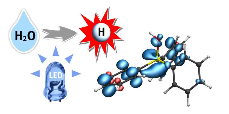
from water (H2O) is transferred to a phosphine-water radical cation under the supply of light energy (LED). This important radical intermediate can further transfer the hydrogen atom (white) to the substrate. The blue regions indicate the electron spin distribution. Credit: Christian Mück-Lichtenfeld

Hydrogen is seen as an energy source of the future—at least, when it is produced in a climate-friendly way. Hydrogen can also be important for the production of active ingredients and other important substances. To produce hydrogen, water (H2O) can be converted into hydrogen gas (H2) by means of a series of chemical processes. However, as water molecules are very stable, splitting them into hydrogen and oxygen presents a big challenge to chemists. For it to succeed at all, the water first has to be activated using a catalyst; then it reacts more easily.

A team of researchers led by Prof. Armido Studer at the Institute of Organic Chemistry at Münster University (Germany) has developed a photocatalytic process in which water, under mild reaction conditions, is activated through triaryl phosphines, and not—as in most other processes—through transition metal complexes.

This strategy, which has now been published in Nature, will open a new door in the highly active field of research relating to radical chemistry, says the team. Radicals are, as a rule, highly reactive intermediates. The team uses a special intermediate—a phosphine-water radical cation—as activated water, from which hydrogen atoms from H2O can be easily split off and transferred to a further substrate. The reaction is driven by light energy.

“Our system,” says Prof. Studer, “offers an ideal platform for investigating unresearched chemical processes which use the hydrogen atom as a reagent in synthesis.”

Dr. Christian Mück-Lichtenfeld, who analyzed the activated water complexes using theoretical methods, says, “The hydrogen-oxygen bond in this intermediate is extraordinarily weak, making it possible to transfer a hydrogen atom to various compounds.”

Dr. Jingjing Zhang, who carried out the experimental work, adds, “The hydrogen atoms of the activated water can be transferred to alkenes and arenes under very mild conditions, in so-called hydrogenation reactions.”

Hydrogenation reactions are enormously important in pharmaceutical research, in the agrochemical industry and in materials sciences.

More information: Jingjing Zhang et al, Photocatalytic phosphine-mediated water activation for radical hydrogenation, Nature (2023). DOI: 10.1038/s41586-023-06141-1

Journal information: Nature 

Provided by University of Münster 

Research students turn Schrödinger’s cat on its head

UW students have turned Schrödinger's cat on its head
Students in the laboratory presenting rotation of Schrödinger cat states. No actual cats were hurt during the project. Credit: S. Kurzyna and B. Niewelt, source: University of Warsaw

Students at the Faculty of Physics of the University of Warsaw (UW) and researchers from the QOT Center for Quantum Optical Technologies have developed an innovative method that allows the fractional Fourier Transform of optical pulses to be performed using quantum memory. This achievement is unique on the global scale, as the team was the first to present an experimental implementation of the said transformation in this type of system.

The results of the research were published in the journal Physical Review Letters. In their work, the students tested the implementation of the fractional Fourier Transform using a double optical pulse, also known as a “Schrödinger’s cat” state.

The spectrum of the pulse and temporal distribution

Waves, such as light, have their own characteristic properties—pulse duration and frequency (corresponding, in the case of light, to its color). It turns out that these characteristics are related to each other through an operation called the Fourier Transform, which makes it possible to switch from describing a wave in time to describing its spectrum in frequencies.

The fractional Fourier Transform is a generalization of the Fourier Transform that allows a partial transition from a description of a wave in time to a description in frequency. Intuitively, it can be understood as a rotation of a distribution (for example, the chronocyclic Wigner function) of the considered signal by a certain angle in the time-frequency domain.

It turns out that transforms of this type are exceptionally useful in the design of special spectral-temporal filters to eliminate noise and enable the creation of algorithms that make it possible to use the quantum nature of light to distinguish pulses of different frequencies more precisely than traditional methods. This is especially important in spectroscopy, which helps study the chemical properties of matter, and telecommunications, which requires the transmission and processing of information with high precision and speed.

An ordinary glass lens is capable of focusing a monochromatic beam of light falling on it to almost a single point (focus). Changing the angle of incidence of light on the lens results in a change in the position of the focus. This allows us to convert angles of incidence into positions, obtaining the analogy of the Fourier Transform, in the space of directions and positions. A classical spectrometer based on a diffraction grating uses this effect to convert the wavelength information of light into positions, allowing us to distinguish between spectral lines.

Time and frequency lenses

Similarly to the glass lens, time and frequency lenses allow the conversion of a pulse’s duration into its spectral distribution, or effectively, perform a Fourier transform in time and frequency space. The right selection of powers of such lenses makes it possible to perform a fractional Fourier Transform. In the case of optical pulses, the action of time and frequency lenses corresponds to applying quadratic phases to the signal.

To process the signal, the researchers used a quantum memory—or more precisely a memory equipped with quantum light processing capabilities—based on a cloud of rubidium atoms placed in a magneto-optical trap. The atoms were cooled to a temperature of tens of millions of degrees above absolute zero. The memory was placed in a changing magnetic field, allowing components of different frequencies to be stored in different parts of the cloud. The pulse was subjected to a time lens during writing and reading, and a frequency lens acted on it during storage.

The device developed at the UW allows the implementation of such lenses over a very wide range of parameters and in a programmable way. A double pulses is very prone to decoherence, hence it is often compared to the famous Schrödinger cat—a macroscopic superposition of being dead and alive, almost impossible to achieve experimentally. Still, the team was able to implement faithful operations on those fragile dual-pulse states.

Before direct application in telecommunications, the method must first be mapped to other wavelengths and parameter ranges. Fractional Fourier transform, however, could prove crucial for optical receivers in state-of-the-art networks, including optical satellite links. A quantum light processor developed at the UW makes it possible to find and test such new protocols in an efficient way.

More information: Bartosz Niewelt et al, Experimental Implementation of the Optical Fractional Fourier Transform in the Time-Frequency Domain, Physical Review Letters (2023). DOI: 10.1103/PhysRevLett.130.240801

Journal information: Physical Review Letters 

Provided by University of Warsaw 

New silica-based adsorbent developed for selective separation of radioactive strontium from acidic medium

by Zhang Nannan, Chinese Academy of Sciences

New silica-based adsorbent developed for selective separation of radioactive strontium from acidic medium
Schematic illustration of SbCl3/SiO2 and Sb2O5/SiO2 preparation processes. Credit: Zhang Shichang

Prof. Huang Qunying’s team from the Hefei Institutes of Physical Science of the Chinese Academy of Sciences has developed a novel inorganic silica-based adsorbent for the highly selective separation of strontium from acidic medium. The results were published in Separation and Purification Technology.

Radioactive strontium (90Sr) is considered to be one of the most dangerous radionuclides due to its high biochemical toxicity. During the vitrification process of high-level liquid waste, the presence of 90Sr can cause instability of the vitrification substrate, resulting in radionuclide leaching. Removal of 90Sr can reduce the heat generation and shorten the cooling time of the vitrification substrate in the repository, which is favorable for further deep geological disposal of the radioactive waste.

To address the above issues, Prof. Huang’s team developed a novel silica-based adsorbent Sb2O5/SiO2 by a two-step method, i.e., vacuum impregnation followed by oxidation, and investigated the adsorption behavior of the adsorbent on strontium stable nuclide in low and high acid mediums (i.e., pH 6 and 1 M HNO3).

New silica-based adsorbent developed for selective separation of radioactive strontium from acidic medium
Adsorption mechanism and DFT calculations: (a) 
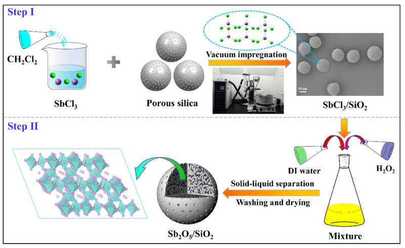
Schematic illustration of the adsorption mechanism; (b, c) Charge density difference of Sb2O5 before and after adsorbed Sr. Credit: Zhang Shichang

The experimental results showed that the prepared adsorbent possessed good acid resistance stability and exhibited favorable adsorption on strontium stable nuclide in both low and high acid mediums. The mechanistic results revealed that the adsorption mechanism was ion exchange, and the adsorption was accompanied by charge transfer and reduction of adsorption energy.

This study not only develops a novel method for the preparation of highly stable silica-based adsorbent, but also provides relevant experimental data and theoretical basis for the selective separation of strontium in acidic environments.

More information: Shichang Zhang et al, Efficient separation of strontium in different environments with novel acid-resistant silica-based ion exchanger, Separation and Purification Technology (2023). DOI: 10.1016/j.seppur.2023.124347

Provided by Chinese Academy of Sciences 

Novel insights on the interplay of electromagnetism and the weak nuclear force

New insights on the interplay of electromagnetism and the weak nuclear force
A spinning neutron disintegrates into a proton, electron, and antineutrino when a down quark in the neutron emits a W boson and converts into an up quark. The exchange of quanta of light (γ) among charged particles changes the strength of this transition. Credit: Vincenzo Cirigliano, Institute for Nuclear Theory

Outside atomic nuclei, neutrons are unstable particles, with a lifetime of about fifteen minutes. The neutron disintegrates due to the weak nuclear force, leaving behind a proton, an electron, and an antineutrino. The weak nuclear force is one of the four fundamental forces in the universe, along with the strong force, the electromagnetic force, and the gravitational force.

Comparing experimental measurements of neutron decay with theoretical predictions based on the weak nuclear force can reveal as-yet undiscovered interactions. To do so, researchers must achieve extremely high levels of precision. A team of nuclear theorists has uncovered a new, relatively large effect in neutron decay that arises from the interplay of the weak and electromagnetic forces.

This research identified a shift in the strength with which a spinning neutron experiences the weak nuclear force. This has two major implications. First, scientists have known since 1956 that due to the weak force, a system and one built like its mirror image do not behave in the same way. In other words, mirror reflection symmetry is broken. This research affects the search for new interactions, technically known as “right-handed currents,” that, at very short distances of less than one hundred quadrillionths of a centimeter, restore the universe’s mirror-reflection symmetry. Second, this research points to the need to compute electromagnetic effects with higher precision. Doing so will require the use of future high-performance computers.

A team of researchers computed the impact of electromagnetic interactions on neutron decay due to the emission and absorption of photons, the quanta of light. The team included nuclear theorists from the Institute for Nuclear Theory at the University of Washington, North Carolina State University, the University of Amsterdam, Los Alamos National Laboratory, and Lawrence Berkeley National Laboratory and their results have been published in Physical Review Letters.

The calculation was performed with a modern method, known as “effective field theory,” that efficiently organizes the importance of fundamental interactions in phenomena involving strongly interacting particles. The team identified a new percent-level shift to the nucleon axial coupling, gA, which governs the strength of decay of a spinning neutron. The new correction originates from the emission and absorption of electrically charged pions, which are mediators of the strong nuclear force. While effective field theory provides an estimate of the uncertainties, improving on the current precision will require advanced calculations on Department of Energy supercomputers.

The researchers also assessed the impact on searches of right-handed current. They found that after including the new correction, experimental data and theory are in good agreement and current uncertainties still allow for new physics at a relatively low mass scale.

More information: Vincenzo Cirigliano et al, Pion-Induced Radiative Corrections to Neutron β Decay, Physical Review Letters (2022). DOI: 10.1103/PhysRevLett.129.121801

Journal information: Physical Review Letters 

Provided by US Department of Energy 

Researchers develop method to simplify one step of radioisotope production

by Kristi L. Bumpus, Oak Ridge National Laboratory

Faster, safer target prep
Illustration shows how the composite is pressed into a seamless aluminum liner, which is then sealed with an aluminum powder cap. Credit: Chris Orosco/ORNL, U.S. Dept. of Energy

Oak Ridge National Laboratory researchers have developed a method to simplify one step of radioisotope production—and it’s faster and safer.

ORNL produces several radionuclides from irradiated radium-226 targets, including actinium-227 and thorium-228, both used in cancer treatments. Continuously improving isotopes for human health is one of the lab’s missions.

Currently, it takes workers two weeks to prepare radium-226 targets for irradiation in the High Flux Isotope Reactor. The targets are exposed to radiation throughout the process, which involves pressing radium carbonate aluminum composite into 10 pellets—one each day—and sealing them into an aluminum capsule.

The new method uses a single, seamless aluminum liner with aluminum powder caps to press and seal the radium carbonate. This minimizes the time required to prepare targets, significantly decreasing radiation doses to workers, and also reduces the targets’ failure rate.

Provided by Oak Ridge National Laboratory 

Chemists develop new method to create chiral structures

by University of California – Riverside

Chemists develop new method to create chiral structures
The photos depict the vibrant colors exhibited by a dispersion of magnetic nanoparticles when subjected to magnetic fields with varying chiral distributions, as observed through polarized lenses. Credit: Yin lab, UC Riverside.

Some molecules exist in two forms, such that their structures and their mirror images are not superimposable, like our left and right hands. Called chirality, it is a property these molecules have due to their asymmetry. Chiral molecules tend to be optically active because of how they interact with light. Oftentimes, only one form of a chiral molecule exists in nature, for example, DNA. Interestingly, if a chiral molecule works well as a drug, its mirror image could be ineffective for therapy.

In trying to produce artificial chirality in the lab, a team led by chemists at the University of California, Riverside, has found that the distribution of a magnetic field is itself chiral. The research paper titled, “A magnetic assembly approach to chiral superstructures,” appears in the journal Science.

“We discovered that the magnetic field lines produced by any magnet, including a bar magnet, have chirality,” said Yadong Yin, a professor of chemistry, who led the team. “Further, we were also able to use the chiral distribution of the magnetic field to coax nanoparticles into forming chiral structures.”

Traditionally, researchers have used “templating” to create a chiral molecule. A chiral molecule is first used as the template. Achiral (or non-chiral) nanoparticles are then assembled on this template, allowing them to mimic the structure of the chiral molecule. The drawback to this technique is that it cannot be universally applied, being heavily dependent on the specific composition of the template molecule. Another shortcoming is the newly formed chiral structure cannot be easily positioned at a specific location on, say, an electronic device.

“But to gain an optical effect, you need a chiral molecule to occupy a particular place on the device,” Yin said. “Our technique overcomes these drawbacks. We are able to rapidly form chiral structures by magnetically assembling materials of any chemical composition at scales ranging from molecules to nano- and microstructures.”

Yin explained that his team’s method uses permanent magnets that consistently rotate in space to generate the chirality. He said transferring chirality to achiral molecules is done by doping, that is incorporating guest species, such as metals, polymers, semiconductors, and dyes into the magnetic nanoparticles used to induce chirality.

Yin said chiral materials acquire an optical effect when they interact with polarized light. In polarized light, light waves vibrate in a single plane, reducing the overall intensity of the light. As a result, polarized lenses in sunglasses cut glare to our eyes, while non-polarized lenses do not.

“If we change the magnetic field that produces a material’s chiral structure, we can change the chirality, which then creates different colors that can be observed through the polarized lenses,” Yin said. “This color change is instantaneous. Chirality can also be made to disappear instantaneously with our method, allowing for rapid chirality tuning.”

The findings could have applications in anti-counterfeit technology. A chiral pattern that signifies the authenticity of an object or document would be invisible to the naked eye but visible when seen through polarized lenses. Other applications of the findings are in sensing and the field of optoelectronics.

“More sophisticated optoelectronic devices can be made by taking advantage of the tunability of chirality that our method allows,” said Zhiwei Li, the first author of the paper and former graduate student in Yin’s lab. “Where sensing is concerned, our method can be used to rapidly detect chiral or achiral molecules linked to certain diseases, such as cancer and viral infections.”

Yin and Li were joined in the research by a team of graduate students in Yin’s lab, including Qingsong Fan, Zuyang Ye, Chaolumen Wu, and Zhongxiang Wang. Li is now a postdoctoral researcher at Northwestern University in Illinois.

More information: Zhiwei Li et al, A magnetic assembly approach to chiral superstructures, Science (2023). DOI: 10.1126/science.adg2657www.science.org/doi/10.1126/science.adg2657

Journal information: Science 

Provided by University of California – Riverside 

New spectroscopy method reveals accelerated relaxation dynamics in compressed cerium-based metallic glass

Accelerated relaxation dynamics in compressed cerium-based metallic glass
Two-time correlation functions of the ce-based MG measured by HP-XPCS at different pressures during compression. At each pressure, the width of the reddish diagonal contour is proportional to the relaxation time, which broadens below 2.9 GPa and then narrows during further compression. Credit: Dr. Qiaoshi Zeng of HPSTAR

A major stumbling block in our understanding of glass and glass phenomena is the elusive relationship between relaxation dynamics and glass structure. A team led by Dr. Qiaoshi Zeng from HPSTAR recently developed a new in situ high-pressure wide-angle X-ray photon correlation spectroscopy method to enable atomic-scale relaxation dynamics studies in metallic glass systems under extreme pressures. The study is published in Proceedings of the National Academy of Sciences (PNAS).

Metallic glasses (MGs), with many superior properties to both conventional metals and glasses, have been the focus of worldwide research. As thermodynamically metastable materials, like typical glasses, MGs spontaneously evolve into their more stable states all the time through various relaxation dynamic behaviors.

These relaxation behaviors have significant effects on the physical properties of MGs. Still, until now, scientists’ ability to deepen the understanding of glass relaxation dynamics and especially its relationships with atomic structures has been limited by the available techniques.

“Thanks to the recent improvements in synchrotron X-ray photon correlation spectroscopy (XPCS), measuring the collective particle motions of glassy samples with a high resolution and broad coverage in the time scale is possible, and thus, various microscopic dynamic processes otherwise inaccessible have been explored in glasses,” said Dr. Zeng.

“However, the change in atomic structures is subtle in previous relaxation process measurements, which makes it still difficult to probe the relationship between the structure and relaxation behavior. To overcome this problem, we decided to employ high pressure because it can effectively alternate the structure of various materials, including MG.”

To this end, the team developed in situ high-pressure synchrotron wide-angle XPCS to probe a cerium-based MG material during compression. In situ high-pressure wide-angle XPCS revealed that the collective atomic motion initially slows down, as generally expected with increasing density. Then, counter-intuitively it accelerates with further compression, showing an unusual non-monotonic pressure-induced steady relaxation dynamics crossover at ~3 GPa.

Furthermore, by combining these results with in situ high-pressure synchrotron X-ray diffraction, the relaxation dynamics anomaly closely correlates with the dramatic changes in local atomic structures during compression, rather than monotonically scaling with either the sample density or overall stress level.

“With density increases, atoms in glasses generally get more difficult to move or diffuse, slowing down its relaxation dynamics. This is what we normally expect from hydrostatic compression,” Dr. Zeng explained.

“So the non-monotonic relaxation behavior observed here in the cerium-based MG under pressure is quite unusual, which indicates besides density, structural details could also play an important role in glass relaxation dynamics,” Dr. Zeng explained.

These findings demonstrate that there is a close relationship between glass relaxation dynamics and atomic structures in MGs. The technique Dr. Qiaoshi Zeng’s group developed here can also be extended to explore the relationship between relaxation dynamics and atomic structures in various glasses, especially those significantly tunable by compression, offering new opportunities for glass relaxation dynamics studies at extreme conditions.

More information: Qiaoshi Zeng et al, Pressure-induced nonmonotonic cross-over of steady relaxation dynamics in a metallic glass, Proceedings of the National Academy of Sciences (2023). DOI: 10.1073/pnas.230228112

Journal information: Proceedings of the National Academy of Sciences 

Provided by Center for High Pressure Science & Technology Advanced Research